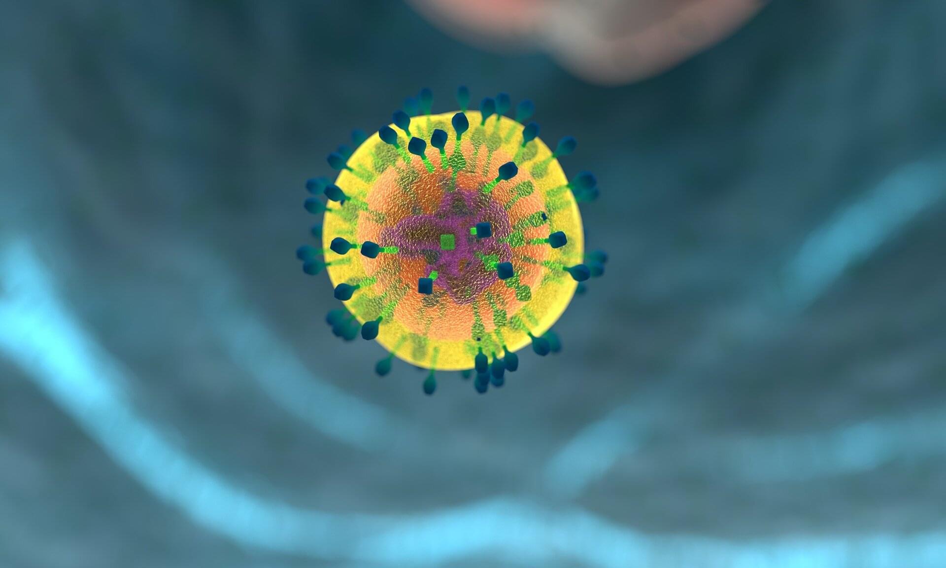
Researchers have uncovered how the brain’s immune cells, called microglia, can act as protectors rather than destroyers in Alzheimer’s disease.
Motorized AI webcam makes life feel dynamic again.
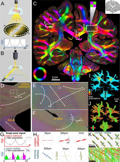
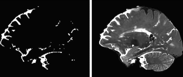
To understand brain diseases, neuroscientists try to understand the intricate maze of nerve fibers in our brains. For analysis under a microscope, brain tissue is often immersed in paraffin wax to create high-quality slices. But until now, it has been impossible to precisely trace the densely packed nerves in these slices. Researchers from Delft, Stanford, Jülich, and Rotterdam have achieved a milestone: using the ComSLI technique, they can now map the fibers in any tissue sample with micrometer precision. The research is published in Nature Communications.
Micron-resolution fiber mapping in histology independent of sample preparation.
Georgiadis and colleagues conduct micron-resolution fibre mapping on multiple histological tissue sections. Their light-scattering technique works across different sample preparations and tissue types, including formalin-fixed paraffin-embedded brain sections.
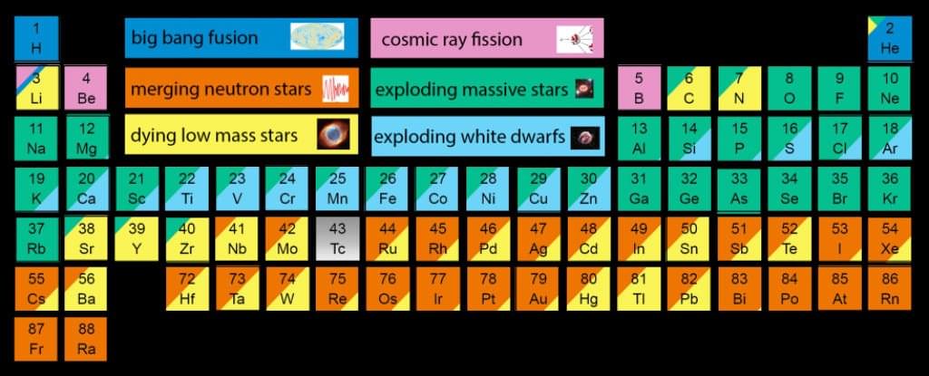
Here’s something to think about: the average adult human is made up of-1 (7 octillion) atoms, and most of them are hydrogen — the most common element in the Universe, produced by the Big Bang 13.8 billion years ago.
The rest of those atoms were forged by ancient stars merging and exploding billions of years after the formation of the Universe, and a tiny amount can be attributed to cosmic rays — high-energy radiation that mostly originates from somewhere outside the Solar System.
As astronomer Carl Sagan once said in an episode of Cosmos, “The nitrogen in our DNA, the calcium in our teeth, the iron in our blood, the carbon in our apple pies were made in the interiors of collapsing stars. We are made of starstuff.”
Scientists have developed a nanoparticle-based treatment that successfully reversed Alzheimer’s disease in mice.
As detailed in a new paper published in the journal Signal Transduction and Targeted Therapy, the team co-led by the Institute for Bioengineering of Catalonia, Spain (IBEC), and West China Hospital, Sichuan University, developed bioactive “supramolecular drugs” that can proactively repair the blood-brain barrier.
The barrier plays an important role in the health of the brain, defending it from harmful substances and other pathogens. Alzheimer’s has been linked to a weakening of the barrier’s integrity, allowing for impairing toxins to make it through.

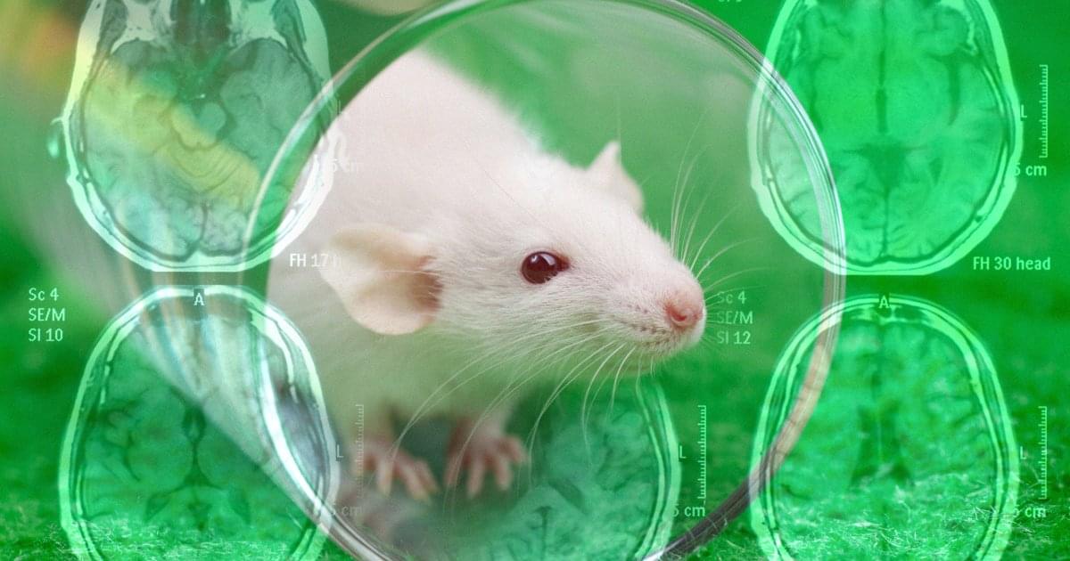
Even small amounts of bisphenol A can lead to long-term health effects. When researchers studied adult rats exposed in the fetal stage, they found that females had developed a more masculine and males a more feminine gene expression pattern. This led to females progressing towards a cancer-like state, while males progressed towards metabolic syndrome, which can increase the risk of diabetes and heart disease.
Bisphenol A is a synthetic chemical with estrogen-like properties that is commonly used in food packaging materials. The substance is banned in many products, but is still present in some packaging. Levels of bisphenol A in people’s bodies are often above levels considered safe, with previous research showing that the substance can cause adverse health effects.
Females masculinized and males feminized In the current study, published in Communications Medicine, researchers investigated how bisphenol A affects the body during the fetal stage.
Chimeric Antigen Receptor (CAR) T cell therapies have revolutionized cancer treatment—but so far, their success has been largely limited to blood cancers. Solid tumors, which account for around 90% of all adult cancers, remain a major challenge because they are difficult for CAR T cells to infiltrate and are often highly heterogeneous, making them harder to target with a single therapy.
Researchers at Monash University, in collaboration with scientists from the Peter MacCallum Cancer Center, used CRISPR-based gene editing or a PTPN2 inhibitor to enhance the function of human CAR T cells engineered to recognize an antigen expressed on many solid tumors.
The study, led by Professor Tony Tiganis and Dr. Florian Wiede, was published in Science Translational Medicine.
Every cell in the body has the same DNA, but different cell types—such as muscle or brain cells—use different parts of it. Transcription factors help cells activate specific genes by reading certain DNA sequences, but since these sequences are common across the genome, scientists have long wondered how the factors know exactly where to bind.
Researchers in the Schübeler lab set out to address this question by looking at two closely related transcription factors—NGN2 and MyoD1—that steer cells toward becoming neurons and muscle cells, respectively. Using stem cells, they switched these transcription factors on one at a time and watched where they attached to the DNA and how they influenced gene expression. Their research is published in the journal Molecular Cell.
They found that the binding of transcription factors to the DNA molecule depends not only on the DNA sequence but also on how open the DNA is and which partner proteins are present. Sometimes, transcription factors act as “pioneer factors” and are able to open tightly packed DNA at specific sites to turn on genes. Small DNA changes—sometimes just one letter—and the proteins these factors partner with can affect whether genes are activated.
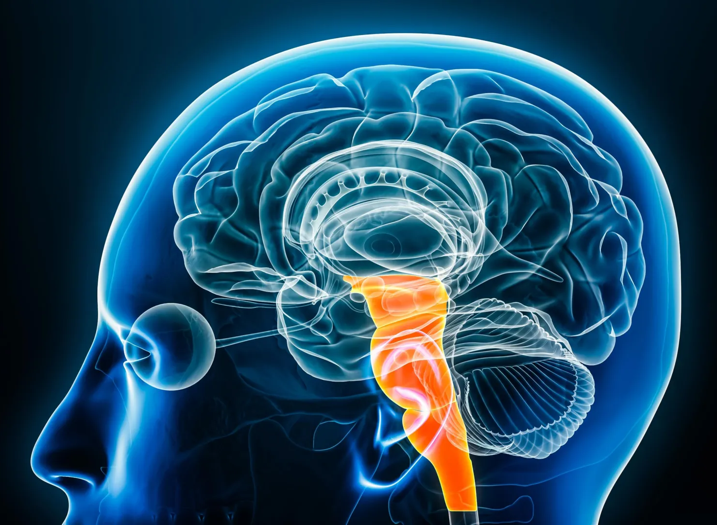
Using powerful 7-Tesla brain imaging, researchers mapped how the brainstem manages pain differently across the body. They discovered that distinct regions activate for facial versus limb pain, showing the brain’s built-in precision pain control system. The findings could lead to targeted, non-opioid treatments that use cannabinoid mechanisms instead of opioids, offering safer pain relief options.