Toggle light / dark theme

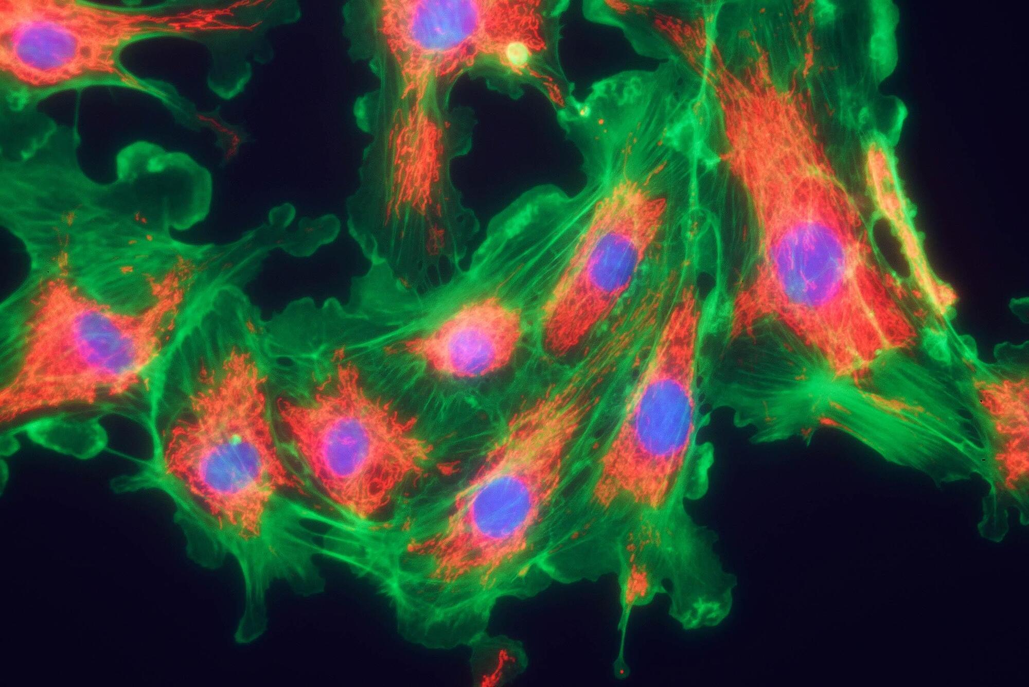
Rochester researchers harnessed adaptive optics to gain insight into the complex workings of the retina and its role in processing color. They have identified elusive retinal ganglion cells (RCGs) in the eye’s fovea that could explain how humans see red, green, blue, and yellow.

Scientists have long wondered how the eye’s three cone photoreceptor types work together to allow humans to perceive color. In a new study in the Journal of Neuroscience, researchers at the University of Rochester used adaptive optics to identify rare retinal ganglion cells (RGCs) that could help fill in the gaps in existing theories of color perception.

The retina has three types of cones to detect color that are sensitive to either short, medium, or long wavelengths of light. Retinal ganglion cells transmit input from these cones to the central nervous system.

Researchers show that the possible cause of local bone erosion in cholesteatomas are fibroblasts from the bone that express a protein called activin A.

Chronic inflammation of the middle ear can cause several problems and complications that can affect a person’s hearing and balance. One such problem is the formation of a cholesteatoma, which is an abnormal collection of cells in the ear that can cause bone erosion if left untreated. In turn, this can cause symptoms such as hearing loss, dizziness, facial paralysis, and even a brain infection.

In a study published in the journal Nature Communications, researchers from Osaka University have revealed the cause of cholesteatomas, which may help in developing new therapies for patients who are suffering from this disease.

News: Skin injuries can modify the gut microbiome, according to a NIAMSfunded study led by UC San Diego.

Read the OpenAccess paper via Nature Portfolio.


The microbial community in the intestine can affect other organs such as the skin but it is not clear if the opposite can occur. Here the authors show that skin wounding affects the microbial composition of the intestinal flora which then enhances DSS induced colitis and intestinal inflammation.

Carbon nanotubes are one of the most elastically strong materials out there.


When I was a kid, I used to take allowance money and occasionally buy rubber-band-powered balsa wood airplanes at a local store. Maybe you’ve seen these. You wind up the rubber band, which stretches the elastomer and stores energy in the elastic strain of the polymer, as in Hooke’s Law (though I suspect the rubber band goes well beyond the linear regime when it’s really wound up, because of the higher order twisting that happens). Rhett Alain wrote about how well you can store energy like this. It turns out that the stored energy per mass of the rubber band can get pretty substantial.

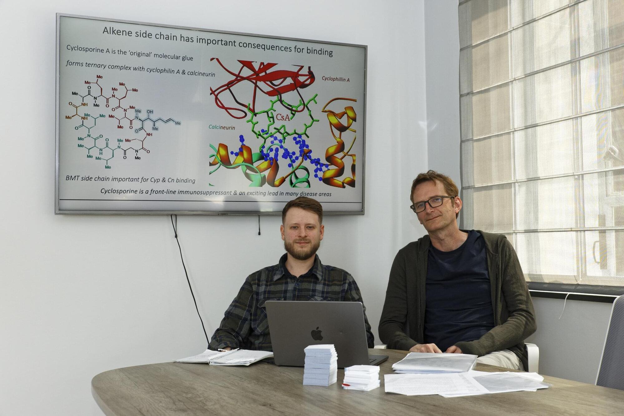
Carbon nanotubes are one of the most elastically strong materials out there. A bit over a decade ago, a group at Michigan State did a serious theoretical analysis of how much energy you could store in a twisted yarn made from single-walled carbon nanotubes. They found that the specific energy storage could get as large as several MJ/kg, as much as four times what you get with lithium ion batteries!

Now, a group in Japan has actually put this to the test, in this Nature Nano paper. They get up to 2.1 MJ/kg, over the lithium ion battery mark, and the specific power (when they release the energy) at about \(10^{6}\) W/kg is not too far away from non-cyclable energy storage media, like TNT. Very cool!

Since the discovery of quantum mechanics more than a hundred years ago, it has been known that electrons in molecules can be coupled to the motion of the atoms that make up the molecules. Often referred to as molecular vibrations, the motion of atoms act like tiny springs, undergoing periodic motion. For electrons in these systems, being joined to the hip with these vibrations means they are constantly in motion too, dancing to the tune of the atoms, on timescales of a millionth of a billionth of a second.

But all this dancing around leads to a loss of energy and limits the performance of organic molecules in applications like organic light emitting diodes (OLEDs), infrared sensors and fluorescent biomarkers used in the study of cells and for tagging diseases such as cancer cells.

Now, researchers using laser-based spectroscopic techniques have discovered ‘new molecular design rules’ capable of halting this molecular dance. Their results, reported in Nature (“Decoupling excitons from high-frequency vibrations in organic molecules”), revealed crucial design principles that can stop the coupling of electrons to atomic vibrations, in effect shutting down their hectic dancing and propelling the molecules to achieve unparalleled performance.

Inside every plant, animal and human cell are billions of molecular machines. They’re made up of proteins, DNA and other molecules, but no single piece works on its own. Only by seeing how they interact together, across millions of types of combinations, can we start to truly understand life’s processes.

In a paper published in Nature, we introduce AlphaFold 3, a revolutionary model that can predict the structure and interactions of all life’s molecules with unprecedented accuracy. For the interactions of proteins with other molecule types we see at least a 50% improvement compared with existing prediction methods, and for some important categories of interaction we have doubled prediction accuracy.

We hope AlphaFold 3 will help transform our understanding of the biological world and drug discovery. Scientists can access the majority of its capabilities, for free, through our newly launched AlphaFold Server, an easy-to-use research tool. To build on AlphaFold 3’s potential for drug design, Isomorphic Labs is already collaborating with pharmaceutical companies to apply it to real-world drug design challenges and, ultimately, develop new life-changing treatments for patients.

Google DeepMind’s newly launched AlphaFold Server is the most accurate tool in the world for predicting how proteins interact with other molecules throughout the cell. It is a free platform that scientists around the world can use for non-commercial research. With just a few clicks, biologists can harness the power of AlphaFold 3 to model structures composed of proteins, DNA, RNA and a selection of ligands, ions and chemical modifications.

AlphaFold Server will help scientists make novel hypotheses to test in the lab, speeding up workflows and enabling further innovation. Our platform gives researchers an accessible way to generate predictions, regardless of their access to computational resources or their expertise in machine learning.

Experimental protein-structure prediction can take about the length of a PhD and cost hundreds of thousands of dollars. Our previous model, AlphaFold 2, has been used to predict hundreds of millions of structures, which would have taken hundreds of millions of researcher-years at the current rate of experimental structural biology.

AlphaFold 3 model is a Google DeepMind and Isomorphic Labs collaboration.

Links and further reading:
Find out more about AlphaFold 3 at https://blog.google/technology/ai/goo
Read the full paper https://www.nature.com/articles/s4158
Access AlphaFold Server: alphafoldserver.com.

Proteins are the molecular machines that sustain every cell and organism, and knowing what they look like will be critical to untangling how they function normally and malfunction in disease. Now researchers have taken a huge stride toward that goal with the development of new machine learning algorithms that can predict the folded shapes of not only proteins but other biomolecules with unprecedented accuracy.

In a paper published today in Nature, Google DeepMind and its spinoff company Isomorphic Labs announced the latest iteration of their AlphaFold program, AlphaFold3, which can predict the structures of proteins, DNA, RNA, ligands and other biomolecules, either alone or bound together in different embraces. The findings follow the tail of a similar update to another deep learning structure-prediction algorithm, called RoseTTAFold All-Atom, which was published in March in Science.

The findings, published in a study in Developmental Cell, reveal that intestinal smooth muscle originates in embryos and forms by the same process that is a hallmark of creating scar tissue when a wound heals.

The smooth muscle sits inside tiny finger-like projections called villi, which absorb fats—also known as lipids—from foods. Contractions of these smooth muscles squeeze absorbed dietary fats through lymphatic capillaries, called lacteals, which send the fats into the systemic blood circulation to produce energy.