Feb 3, 2024
Next-Gen Mitochondrial Uncouplers: Potential Game Changer for Metabolic Disorders
Posted by Chris Smedley in categories: biotech/medical, life extension
Reverse Aging Revolution
A team of University of Wisconsin–Madison scientists has developed the first 3D-printed brain tissue that can grow and function like typical brain tissue.
It’s an achievement with important implications for scientists studying the brain and working on treatments for a broad range of neurological and neurodevelopmental disorders, such as Alzheimer’s and Parkinson’s disease.
“This could be a hugely powerful model to help us understand how brain cells and parts of the brain communicate in humans,” says Su-Chun Zhang, professor of neuroscience and neurology at UW–Madison’s Waisman Center. “It could change the way we look at stem cell biology, neuroscience, and the pathogenesis of many neurological and psychiatric disorders.”
Washington state is experiencing its first known outbreak of a potentially deadly fungus, according to public health officials.
Four patients in the last month have tested positive for Candida auris, or C. auris, Public Health — Seattle & King County said in a release.
The first case occurred in a patient who had recently been admitted to Kindred Hospital Seattle, which was identified through a proactive screening program.
The rare condition posterior cortical atrophy (PCA) involves strange, troubling issues with vision and spatial awareness – including difficulty judging distances, seeing movement, and recognizing objects – and a new study highlights its close relationship to Alzheimer’s disease in more detail than ever before.
PCA and Alzheimer’s have long been linked with each other, because they share a lot of the same pathological changes in the brain. However, the rarity of PCA has made it hard for researchers to fully assess it in relation to Alzheimer’s.
To address that, an international team of researchers analyzed data on 1,092 individuals with PCA, finding that it was a very strong predictor for Alzheimer’s: in 94 percent of cases, tell-tale Alzheimer’s brain changes were observed, and were most likely contributing to PCA.
Summary: Researchers developed the world’s first 3D-printed brain tissue that grows and behaves similarly to natural brain tissue, marking a significant leap forward for neurological and neurodevelopmental disorder research.
This novel 3D-printing technique uses a horizontal layering approach and a softer bio-ink, allowing neurons to interconnect and form networks akin to human brain structures.
The ability to precisely control cell types and arrangements provides unparalleled opportunities to study brain functions and disorders in a controlled environment, offering new avenues for drug testing and understanding brain development and diseases like Alzheimer’s and Parkinson’s.
Heartbeat-induced pulsations of cerebral blood vessels can directly entrain neuronal activity.
An AI algorithm outperformed other screening methods in identifying cervical precancer. The approach could be especially valuable in low-resource settings.
A team of molecular engineers have developed a type of plastic that can be shape-shifted using tempering. In their paper published in the journal Science the team, from the University of Chicago, with a colleagues from the US DEVCOM Army Research Laboratory, Aberdeen Proving Ground, the National Institutes of Standards and Technology and the NASA Glenn Research Center, describe how they made their plastic and how well it was able to shape shift when they applied various types of tempering.
Haley McAllister and Julia Kalow, with Northwestern University, have published a Perspective piece in the same issue of Science outlining the work.
Over the past several years, it has become evident that the use of plastics in products is harmful to not only the environment but also human health —bits of plastic have been found in the soil, the atmosphere, the oceans, and the human body.
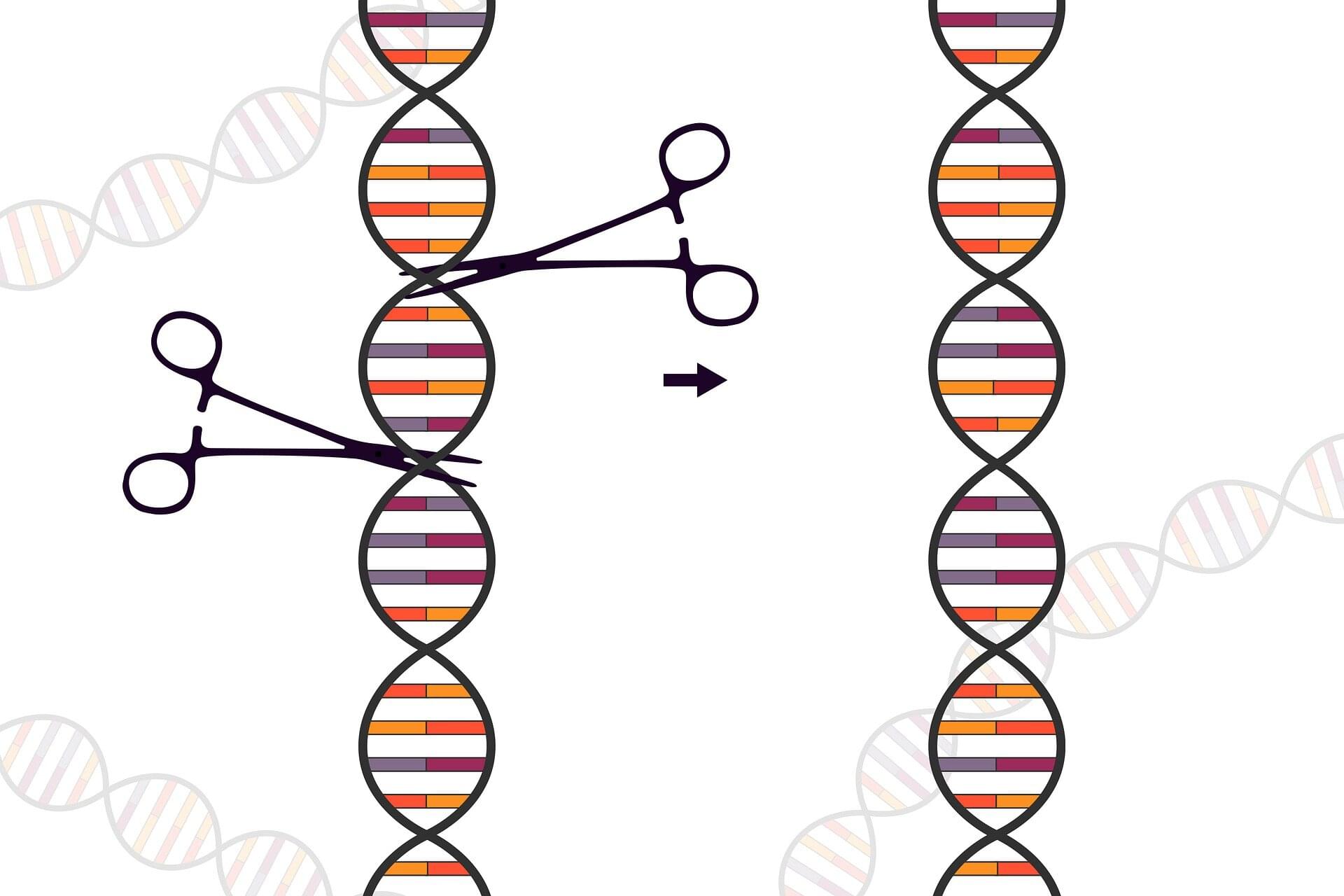
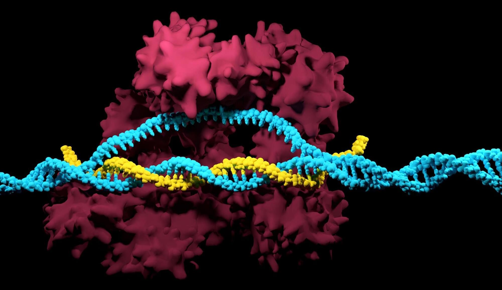
Some hereditary genetic defects cause an exaggerated immune response that can be fatal. Using the CRISPR-Cas9 gene-editing tool, such defects can be corrected, thus normalizing the immune response, as researchers led by Klaus Rajewsky from the Max Delbrück Center now report in Science Immunology.
Familial hemophagocytic lymphohistiocytosis (FHL) is a rare disease of the immune system that usually occurs in infants and young children under the age of 18 months. The condition is severe and has a high mortality rate. It is caused by various gene mutations that prevent cytotoxic T cells from functioning normally. These are a group of immune cells that kill virus–infected cells or otherwise altered cells.
If a child with FHL contracts a virus—such as the Epstein-Barr virus (EBV), but also other viruses—the cytotoxic T cells cannot eliminate the infected cells. Instead, the immune response gets out of control. This leads to a cytokine storm and an excessive inflammatory reaction that affects the entire organism.
Plant cell-surface receptors that are known to participate in immunity, development, and reproductive processes include the LRR-, G-lectin-, Wall-associated kinase (WAK)-, Domain of Unknown Function 26 (Duf26)-, L-lectin-, Lysin motif (LysM)-, and Malectin-containing RLKs and RLPs (Fig. 1a–h). There are additional RLK families with different ectodomains, such as the proline-rich extensin-like receptor kinases (PERKs) and thaumatin-like protein kinases (TLPKs)9,13. However, their function in immunity is not well-characterized. Cell-surface receptors with LRR-, G-lectin-, WAK-, and LysM-ectodomains have been reported to recognise PAMPs, while others perceive self-molecules or unidentified ligands (Fig. 1h; Supplementary Fig. 1). Recognition of the diverse array of ligands is likely to be accomplished by variable structures and combinations of different ectodomains (Fig. 1a–g). To trace the origins of different receptor classes within the plant lineage, we first identified RLKs and RLPs in 350 genomes from Glaucophyta, red algae, green algae, Bryophytes, and Tracheophytes. We define here RLKs as any proteins with both 1–2 TMs and KDs, and RLPs as any protein with 1–2 TMs, but lack KDs. In total, we identified 177,645 RLKs, almost up to 70% of which possess either LRR-, G-lectin-, WAK-, Duf26-, L-lectin-, LysM-and Malectin-ectodomains (Fig. 1i). Next, we searched for proteins with these ectodomains and TMs that lack KDs and found 41,144 RLPs (Fig. 1j). We further examined which of the identified RLKs and RLPs families are likely to be involved in immunity. A previous report suggested a positive correlation between the gene family sizes of cell-surface immune receptors and intracellular immune receptors (the NB-ARC family) across the angiosperms4. We examined the correlation between the relative size (%; number of identified genes in the family/numbers of searched genes × 100; see methods) of the RLK families, the RLPs families, and the NB-ARC family in each genome. Notably, most RLK families (except for the LysM-RLKs) exhibit positive correlations with the NB-ARC family, while most RLP families (except for the LRR-RLPs) do not exhibit positive correlation with the NB-ARC family (Main Fig. 1k). Furthermore, we checked the expression level of these receptor families in Arabidopsis thaliana during immunity. Notably, the RLKs, except for LRR-and Malectin-RLKs, generally exhibit higher expression levels compared to the RLPs during immunity (Main Fig. 1k; Supplementary Fig. 2). These data collectively suggest that the RLKs are more likely to be involved in immunity than the RLPs.
Next, we examined the presence or absence of ectodomains (LRR-, G-lectin-, WAK-, Duf26-, L-lectin-, LysM-and Malectin-ectodomains lacking TM or KD; ectodomain-only proteins), RLPs (TM-bound ectodomains) and RLKs (ectodomains encompassing both TM and KD) in the plant lineage (Fig. 2; Supplementary Fig. 3; Supplementary Data 1a–c). Ectodomains exhibit an ancient heritage, with LRR-, WAK-, LysM-, Malectin-, and L-lectin-domains dating back to the era of Glaucophyta. Similarly, relatively ancient counterparts such as LRR-RLPs, WAK-RLPs, LysM-RLPs, Malectin-RLPs, and L-lectin-RLPs are found in both Glaucophyta and Rhodophyta. In contrast, RLKs emerged more recently. Green algae harbour WAK-RLKs, Malectin-RLKs, and G-lectin-RLKs, and LysM-RLKs, L-lectin-RLKs, and Duf-26-RLKs are exclusive to Embryophytes (Fig. 2). Except for LRR-RLPs, all six families of RLP are basal to the RLK families.