It’s never too late to start strengthening your brain.
Category: biotech/medical – Page 2,718
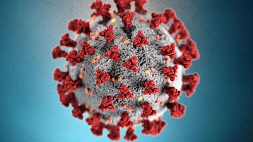
It Takes Two: COVID-19 Coronavirus Possibly ‘Chimera’ of Two Distinct Viruses
;-; They say now it could be a bioweapon possibly like the chimera virus that is why it spread so fast. Many weird reports from god having a battle with the borg to even an alien entity in London but rationally this was a bioweapon as it spread so fast and evolved so fast. Still undecided the origin bit it seems patient zero was in china originally from a bat.
The COVID-19 coronavirus may be a chimera created from the presence of two pre-existing distinct viruses.
Drone Walks A Dog
A man under Coronavirus lockdown used a drone to walk his dog 🐶.
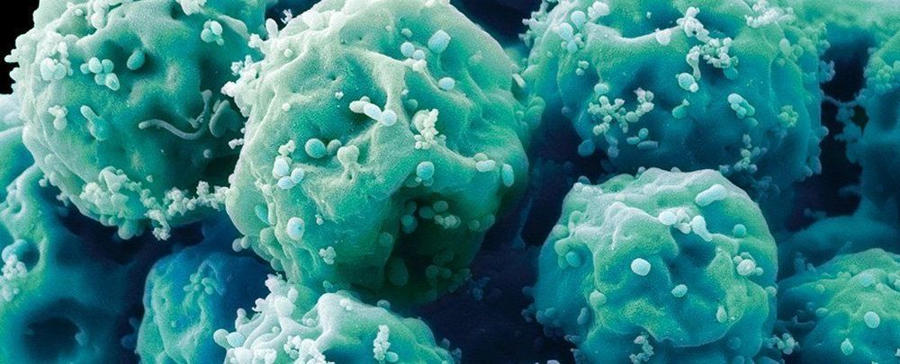
Scientists ‘Reset’ The Age of Stem Cells From a Supercentenarian Who Lived to 114
For the first time, scientists have reprogrammed the stem cells of a 114-year-old woman, the oldest donor to date.
After first transforming cells from her blood sample into induced pluripotent stem cells (iPSCs), the researchers then generated mesenchymal stem cells, which help to maintain and repair tissues like bone, cartilage and fat.
“We set out to answer a big question: Can you reprogram cells this old?” says stem cell biologist Evan Snyder at Sanford Burnham Prebys Medical Discovery Institute in California.

Coronavirus: Nobel Prize winner predicts US will get through crisis sooner than expected
A Nobel Prize winning biology professor has provided a bit of good news amidst the coronavirus gloom; the US may see a downturn in new cases sooner than some models have predicted.
Michael Levitt, a Stanford University biology professor and a 2013 Nobel Prize winner in chemistry, said his models predict the virus is not likely to dwindle on for months or years and – most importantly – is not likely to cause millions of deaths.
Mr Levitt previously predicted – correctly – when China would experience and endure the worst of its coronavirus crisis.
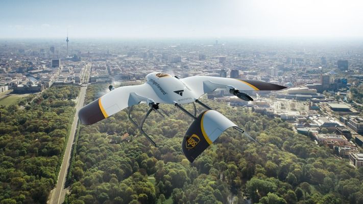
UPS partners with Wingcopter to develop new multipurpose drone delivery fleet
UPS is working with German startup Wingcopter to develop a new type of delivery drone, to be used for the logistics company’s growing commercial drone delivery efforts both in the U.S. and globally. Wingcopter has already designed an electric vertical takeoff and landing (eVTOL) aircraft that has a range of up to 75 miles and can achieve speeds as high as 150 miles per hour in conditions that include wind speeds of up to 45 miles per hour.
Wingcopter will be working closely with UPS’ Flight Forward subsidiary, the dedicated drone delivery unit that UPS developed last year in July to house its commercial drone delivery program. In October, Flight Forward received Federal Aviation Administration (FAA) approval to effectively operate a full-scale “drone airline” at scale for the purpose of package delivery.
Wingcopter has already demonstrated how its drones could operate in commercial settings, including during a demonstration with Merck earlier this year that saw its autonomous eVTOLs carry small packages between the drug company’s various office locations in Darmstadt in Germany. It also used its aircraft to deliver critical medical supplies and life-saving equipment to hard to reach areas, including through partnerships with UNICEF and other relief organizations.
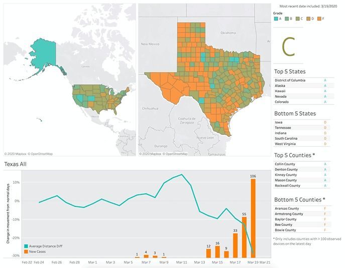
Social Distancing Scoreboard
Social distancing is seen as the most effective way of slowing the spread of the virus causing COVID-19, and is strongly advised by the World Health Organization and the CDC. At Unacast, we wanted to understand which areas of the country are best at exhibiting social distancing behavior. Our hope is that these insights will empower organizations and companies to understand and measure social distancing initiatives and ultimately, save lives.

FDA Grants Experimental Coronavirus Drug Benefits For Rare Disease Treatments
The agency’s decision would provide lucrative incentives to the drug’s maker, Gilead Sciences, and could keep lower-priced generic versions of the medicine off the market for several years if remdesivir is approved for use, public health advocates say.
Remdesivir Gets Rare Disease Perks From FDA : Shots — Health News Gilead Science’s remdesivir, an antiviral medicine being tested for treatment of COVID-19, would get a seven-year monopoly if approved by the Food and Drug Administration.

You could be spreading the coronavirus without realising you’ve got it
Such undocumented cases are still contagious and the study found them to be the source of most of the virus’s spread in China before the restrictions came in. Even though these people were only 55 per cent as contagious as people with symptoms, the study found that they were the source of 79 per cent of further infections, due to there being more of them, and the higher likelihood that they were out and about.
“If somebody’s experiencing mild symptoms, and I think most of us can relate to this, we’re still going to go about our day,” says Shaman. “These people are the major driver of it and they’re the ones who facilitated the spread.”
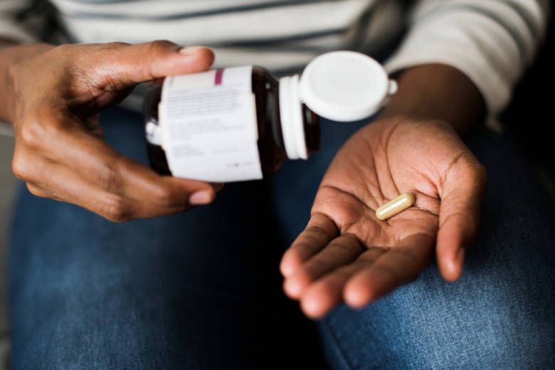
Israelis Invent First Pill to Treat Diabetes Without Insulin Shots
The pill passed second-stage testing with impressive results, placing it on the fast-track for commercial availability, Oramed announced last week, which is music to the ears of the nearly half a billion people worldwide who suffer from diabetes.
An oral insulin capsule passed second stage testing for efficacy and safety, paving the way to diabetes treatments without shots.
By United with Israel Staff
People who suffer from diabetes frequently require insulin shots, causing pain, inconvenience, and visible scars that do not heal. To address this pressing healthcare issue, a company based in Israel developed the first oral insulin capsule, called ORMD-0801, providing diabetes treatment without the need for shots.