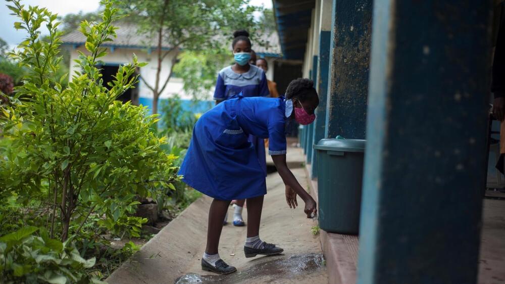
When the results of his study came in, Kondwani Jambo was stunned.
He’s an immunologist in Malawi. And last year he had set out to determine just how many people in his country had been infected with the coronavirus since the pandemic began.
Jambo, who works for the Malawi-Liverpool-Wellcome Trust Clinical Research Programme, knew the total number of cases was going to be higher than the official numbers. But his study revealed that the scale of spread was beyond anything he had anticipated — with a huge majority of Malawians infected long before the omicron variant emerged. “I was very shocked,” he says.
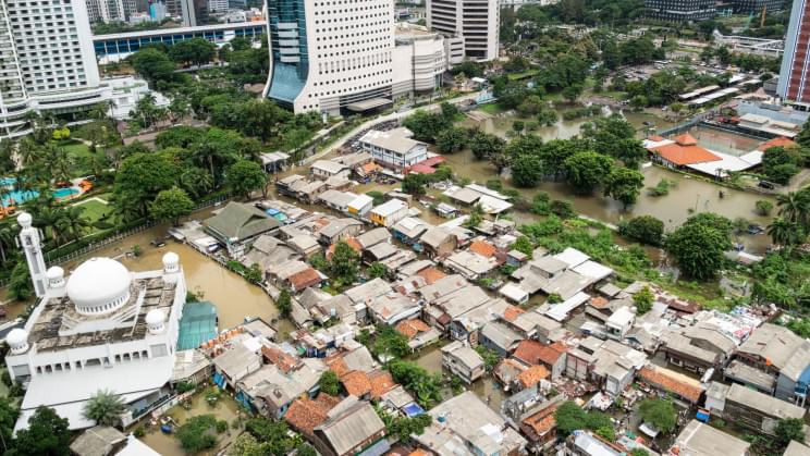
So to fill in the true picture, Jambo and his collaborators turned to another potential source of information: a repository of blood samples that had been collected from Malawians month after month by the national blood bank. And they checked how many of those samples had antibodies for the coronavirus. Their finding: By the start of Malawi’s third COVID-19 wave with the delta variant last summer, as much as 80% of the population had already been infected with some strain of the coronavirus.
New findings from Malawi suggest the country has entered something akin to the endemic stage of the pandemic — along with many other African nations.
