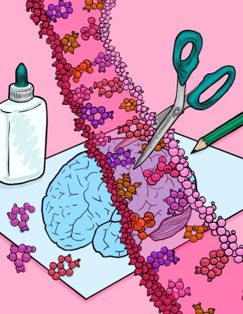
Mount Sinai researchers have cataloged thousands of sites in the brain where RNA is modified throughout the human lifespan in a process known as adenosine-to-inosine (A-to-I) editing, offering important new avenues for understanding the cellular and molecular mechanisms of brain development and how they factor into both health and disease.
In a study published in Cell Reports, the team described how the rate of RNA editing in the brain increases as individuals age, with implications for dissecting the pathology of altered A-to-I editing across a range of neurodevelopmental and aging disorders.
“Our work provides more nuanced and accurate insights into the contribution of RNA modifications by A-to-I editing during human brain development,” says senior author Michael Breen, Ph.D., Assistant Professor of Psychiatry, and Genetics and Genomic Sciences, at the Icahn School of Medicine at Mount Sinai, and a member of the Seaver Center for Autism Research and Treatment.
Reproduction permanently alters females’ bones in ways not previously known, a team of anthropologists has found. Its discovery, based on an analysis of primates, sheds new light on how giving birth can permanently change the body.
“Our findings provide additional evidence of the profound impact that reproduction has on the female organism, further demonstrating that the skeleton is not a static organ, but a dynamic one that changes with life events,” explains Paola Cerrito, who led the research as a doctoral student in NYU’s Department of Anthropology and College of Dentistry.
Specifically, the researchers found that calcium, magnesium, and phosphorus concentrations are lower in females who have experienced reproduction. These changes are linked to giving birth itself and to lactation.
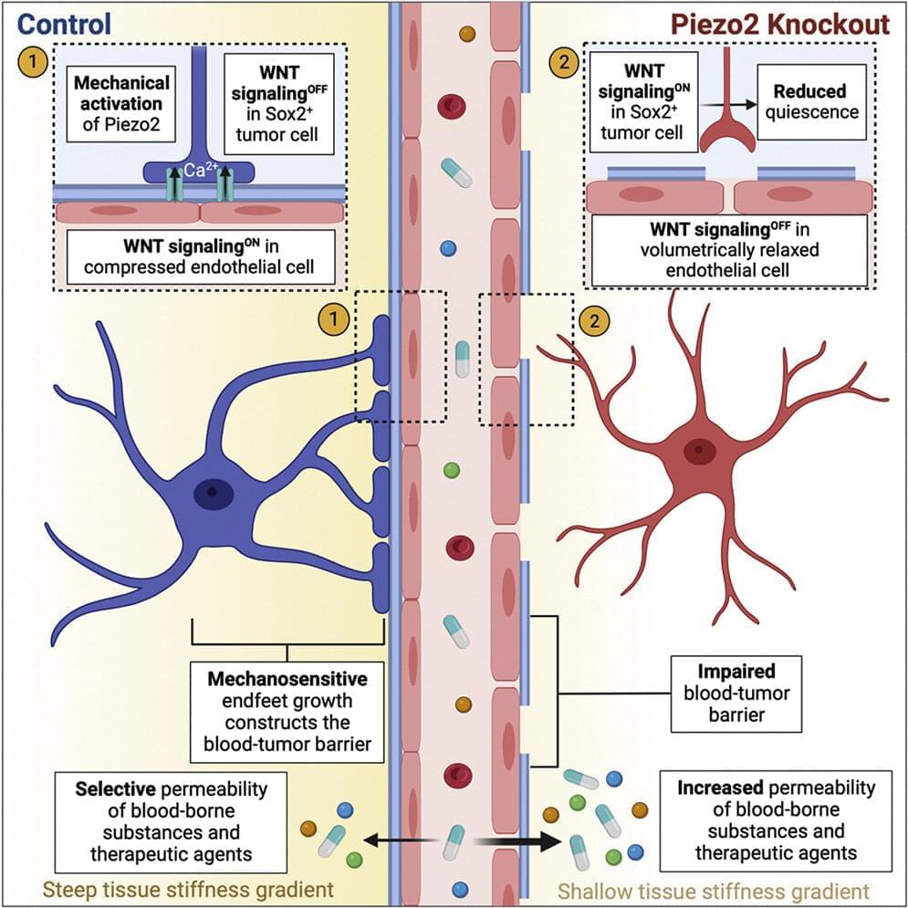
In a new study, scientists have uncovered the mechanics of the blood-tumor barrier, one of the most significant obstacles to improving treatment efficacy and preventing the return of cancerous cells. The research team, led by Dr. Xi Huang, a Senior Scientist in Developmental & Stem Cell Biology program at The Hospital for Sick Children (SickKids), lays the foundation for more effectively treating medulloblastoma, the most common malignant pediatric brain tumor.
“Despite decades of research on brain cancer, the mechanisms that govern the formation and function of the blood-tumor barrier have remained poorly understood,” says Huang, who is also a Principal Investigator at the Arthur and Sonia Labatt Brain Tumor Research Center and Canada Research Chair in Cancer Biophysics. “Our discoveries represent a breakthrough in the understanding of how the blood-tumor barrier forms and works.”
In a paper published today in Neuron, the research team identifies a way to reduce the impact of the blood-tumor barrier on medulloblastoma treatment.
Imagine the booming chords from a pipe organ echoing through the cavernous sanctuary of a massive, stone cathedral.
The sound a cathedral-goer will hear is affected by many factors, including the location of the organ, where the listener is standing, whether any columns, pews, or other obstacles stand between them, what the walls are made of, the locations of windows or doorways, etc. Hearing a sound can help someone envision their environment.
Researchers at MIT and the MIT-IBM Watson AI Lab are exploring the use of spatial acoustic information to help machines better envision their environments, too. They developed a machine-learning model that can capture how any sound in a room will propagate through the space, enabling the model to simulate what a listener would hear at different locations.
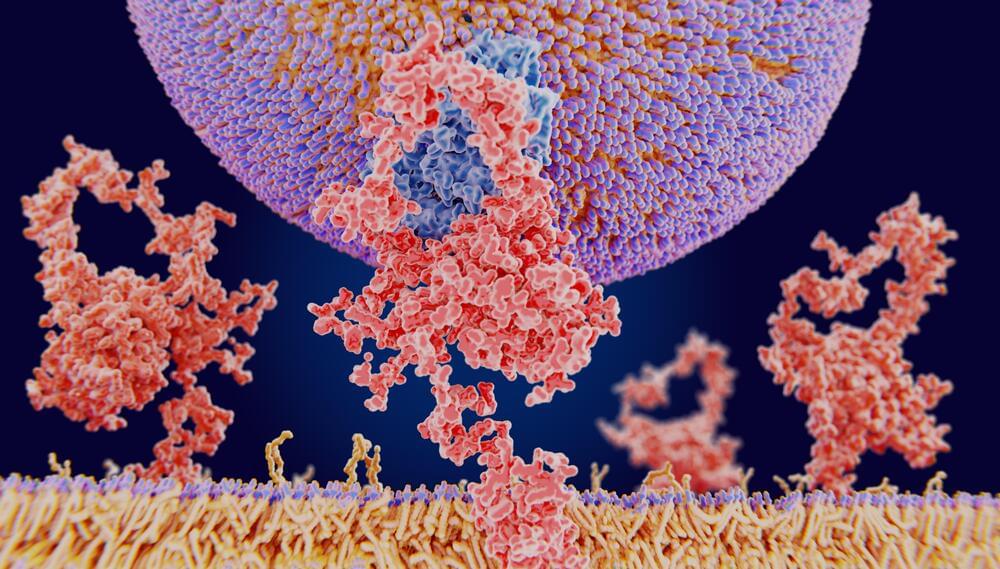
Life on Earth would not exist as we know it, if not for the protein molecules that enable critical processes from photosynthesis and enzymatic degradation to sight and our immune system. And like most facets of the natural world, humanity has only just begun to discover the multitudes of protein types that actually exist. But rather scour the most inhospitable parts of the planet in search of novel microorganisms that might have a new flavor of organic molecule, Meta researchers have developed a first-of-its-kind metagenomic database, the ESM Metagenomic Atlas, that could accelerate existing protein-folding AI performance by 60x.
Metagenomics is just coincidentally named. It is a relatively new, but very real, scientific discipline that studies “the structure and function of entire nucleotide sequences isolated and analyzed from all the organisms (typically microbes) in a bulk sample.” Often used to identify the bacterial communities living on our skin or in the soil, these techniques are similar in function to gas chromatography, wherein you’re trying to identify what’s present in a given sample system.
Similar databases have been launched by the NCBI, the European Bioinformatics Institute, and Joint Genome Institute, and have already cataloged billions of newly uncovered protein shapes. What Meta is bringing to the table is “a new protein-folding approach that harnesses large language models to create the first comprehensive view of the structures of proteins in a metagenomics database at the scale of hundreds of millions of proteins,” according to a Tuesday release from the company. The problem is that, while advances of genomics have revealed the sequences for slews of novel proteins, just knowing what those sequences are doesn’t actually tell us how they fit together into a functioning molecule and going figuring it out experimentally takes anywhere from a few months to a few years. Per molecule. Ain’t nobody got time for that.
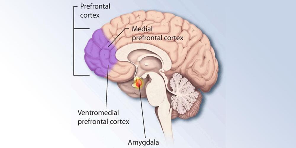
“We were surprised to see that this effect could be seen even with a single dose of nicotine, equivalent to just one cigarette, showing how powerful the effects of smoking are on a woman ’ s brain.”
Emphasizing the preliminary nature of the study and the need for a larger sample, she added: We’re still not sure what the behavioral or cognitive outcomes are; only that nicotine acts on this area of the brain.
However, we note that the affected brain system is a target for addictive drugs, such as nicotine.
A study on a large sample of patients found chronic, long-lasting depression to be associated with reduced brain volume. The reduced volume was found in brain regions relevant for planning one’s behavior, focusing attention, thinking, learning and remembering and also in regions relevant for regulating emotions. The study was published in Neurobiology and Treatment of Depression.
Depression, also called major depressive disorder, is a mood disorder that causes a persistent feeling of sadness and loss of interest. It changes the way a person feels, thinks and behaves. For many people suffering from it, depressive episodes become a recurring event. More than half of patients with depression experience a relapse after 2 years and the probability of recurrent depressive episodes rises to 90% after 3–4 episodes. Studies have indicated that recurring depressive episodes might be linked to structural changes in the brain, but the existing results are not uniform.
Ms. Hannah Lemke and her colleagues analyzed the data of 681 patients from the Marburg-Muenster-Affective-Cohort Study (MACS) in order to better link properties of the course of depressive disorder with specific changes in the brain structure. Patient data were collected at two sites in Germany – Muenster and Marburg.
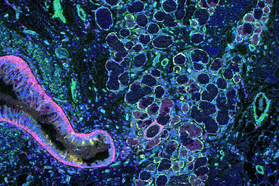
Weill Cornell Medicine researchers have developed a computational method to map the architecture of human tissues in unprecedented detail. Their approach promises to accelerate studies on organ-scale cellular interactions and could enable powerful new diagnostic strategies for a wide range of diseases.
The method, published Oct. 31 in Nature Methods, grew out of the scientists’ frustration with the gap between classical microscopy and modern single-cell molecular analysis. “Looking at tissues under the microscope, you see a bunch of cells that are grouped together spatially—you see that organization in images almost immediately,” said lead author Junbum Kim, a graduate student in physiology and biophysics at Weill Cornell Medicine.
“Now, cell biologists have gained the ability to examine individual cells in tremendous detail, down to which genes each cell is expressing, so they’re focused on the cells instead of focusing on the tissue structure,” he said.