Toggle light / dark theme

De Novo Reconstruction of 3D Human Facial Images from DNA Sequence

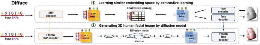
Facial morphology is a distinctive biometric marker, offering invaluable insights into personal identity, especially in forensic science. In the context of high-throughput sequencing, the reconstruction of 3D human facial images from DNA is becoming a revolutionary approach for identifying individuals based on unknown biological specimens. Inspired by artificial intelligence techniques in text-to-image synthesis, it proposes Difface, a multi-modality model designed to reconstruct 3D facial images only from DNA. Specifically, Difface first utilizes a transformer and a spiral convolution network to map high-dimensional Single Nucleotide Polymorphisms and 3D facial images to the same low-dimensional features, respectively, while establishing the association between both modalities in the latent features in a contrastive manner; and then incorporates a diffusion model to reconstruct facial structures from the characteristics of SNPs. Applying Difface to the Han Chinese database with 9,674 paired SNP phenotypes and 3D facial images demonstrates excellent performance in DNA-to-3D image alignment and reconstruction and characterizes the individual genomics. Also, including phenotype information in Difface further improves the quality of 3D reconstruction, i.e. Difface can generate 3D facial images of individuals solely from their DNA data, projecting their appearance at various future ages. This work represents pioneer research in de novo generating human facial images from individual genomics information.

(Repost)


This study has introduced Difface, a de novo multi-modality model to reconstruct 3D facial images from DNA with remarkable precision, by a generative diffusion process and a contrastive learning scheme. Through comprehensive analysis and SNP-FACE matching tasks, Difface demonstrated superior performance in generating accurate facial reconstructions from genetic data. In particularly, Difface could generate/predict 3D facial images of individuals solely from their DNA data at various future ages. Notably, the model’s integration of transformer networks with spiral convolution and diffusion networks has set a new benchmark in the fidelity of generated images to their real images, as evidenced by its outstanding accuracy in critical facial landmarks and diverse facial feature reproduction.

Difface’s novel approach, combining advanced neural network architectures, significantly outperforms existing models in genetic-to-phenotypic facial reconstruction. This superiority is attributed to its unique contrastive learning method of aligning high-dimensional SNP data with 3D facial point clouds in a unified low-dimensional feature space, a process further enhanced by adopting diffusion networks for phenotypic characteristic generation. Such advancements contribute to the model’s exceptional precision and ability to capture the subtle genetic variations influencing facial morphology, a feat less pronounced in previous methodologies.

Despite Difface’s demonstrated strengths, there remain directions for improvement. Addressing these limitations will require a focused effort to increase the model’s robustness and adaptability to diverse datasets. Future research should aim to incorporating variables like age and BMI would allow Difface to simulate age-related changes, enabling the generation of facial images at different life stages an application that holds significant potential in both forensic science and medical diagnostics. Similarly, BMI could help the model account for variations in body composition, improving its ability to generate accurate facial reconstructions across a range of body types.

Imaging-based STAMP technique democratizes single-cell RNA research

Scientists at St. Jude Children’s Research Hospital, the National Center for Genomic Analysis and the University of Adelaide have created a single-cell RNA analysis method that is 47 times cheaper and more scalable than other techniques.

Single-cell RNA sequencing provides scientists with important information about in health and disease. However, the technique is expensive and often prohibits analysis of large numbers of cells.

Scientists from St. Jude Children’s Research Hospital, the National Center for Genomic Analysis and the University of Adelaide have created a method that combines microscopy with single-cell RNA analysis to overcome these limitations. The technique called Single-Cell Transcriptomics Analysis and Multimodal Profiling through Imaging (STAMP) can look at millions of single cells for a fraction of the cost of existing approaches.

Genome of a 28-eyed jellyfish could provide insight on evolution of vision

One of the biggest mysteries of evolution is how species first developed complex vision. Jellyfish are helping scientists solve this puzzle, as the group has independently evolved eyes at least nine separate times. Different species of jellyfish have strikingly different types of vision, from simple eyespots that detect light intensity to sophisticated lens eyes similar to those in humans.

Biologists have studied jellyfish eye structure, light sensitivity, and visual behavior for decades, but the exact genes involved in jellyfish eye formation remain unknown.

Aide Macias-Muñoz, a professor of ecology and , is exploring how eyes and light detection evolved using genetic tools. Her lab has just completed a high-quality genome sequence of Bougainvillia cf. muscus, a small jellyfish-like animal in the Hydrozoa group that has an astonishing 28 eyes.

Biased agonism of GLP-1R and GIPR enhances glucose lowering and weight loss, with dual GLP-1R/GIPR biased agonism yielding greater efficacy

Biased agonism to treat diabetes and obesity.

Agonists of glucagon-like peptide-1 receptor (GLP-1R) and glucose-dependent insulinotropic polypeptide receptor (GIPR) have been used for diabetes and obesity treatment. Mechanism of action and signaling of these receptors are of paramount importance.

The researchers investigate the impact of biased cyclic AMP (cAMP) signaling with a dual GLP-1R/ GIPR agonist.

Biased GLP-1R and GIPR agonism with GLP-1R/GIPR agonist, CT-859 leads to better and prolonged glucose lowering, greater food intake reduction, and weight loss than unbiased agonism.

Biased GIPR agonism synergizes with GLP-1R on food intake suppression and weight loss. https://www.cell.com/cell-reports-medicine/fulltext/S2666&#4…0229-0 https://sciencemission.com/Biased-agonism-of-GLP-1R-and-GIPR


Rodriguez et al. investigate the impact of biased signaling with a dual GLP-1R/GIPR agonist. Biased GLP-1R and GIPR agonism leads to better and prolonged glucose lowering, greater food intake reduction, and weight loss than unbiased agonism. Biased GIPR agonism synergizes with GLP-1R on food intake suppression and weight loss.

Is the Cell’s Antenna Related to Cancer Growth?

Many different types of cells in the body have a tiny projection known as a primary cilium. These cilia act like little signaling hub that can capture information about a cell’s environment and relay it to the cell, ultimately coordinating some cellular responses. The functions of cilia are well known in a few cases, such as in development, where they are crucial to the regulation of certain processes; or in some disorders called ciliopathies, in which genetic mutations lead to ciliary dysfunction and human disease.

Nanoplastics can disrupt gut microbes in mice by interfering with extracellular vesicle-delivered microRNA

Nanoplastics can compromise intestinal integrity in mice by altering the interactions between the gut microbiome and the host, according to a paper in Nature Communications. The study explores the complex interactions of nanoplastics with the gut microenvironment in mice.

Nanoplastics are pieces of plastic less than 1,000 nanometers in diameter, which are created as plastics degrade. Previous research has suggested that uptake can disrupt the gut microbiota; however, the underlying mechanism behind this effect is poorly understood.

Researcher Wei-Hsuan Hsu and colleagues used RNA sequencing, transcriptomic analysis and microbial profiling to analyze the effects of polystyrene nanoplastics on the intestinal microenvironment when ingested in mice. They found that nanoplastic accumulation in the mouse intestine was linked to altered expression of two proteins involved in intestinal barrier integrity (ZO-1 and MUC-13), which could disrupt intestinal permeability.

Discovery of two new genetic disorders improves diagnoses for patients with neurodevelopmental conditions

The discovery of two new genetic disorders comes from a study delivered through the National Institute for Health and Care Research (NIHR) Manchester Biomedical Research Center (BRC) and The University of Manchester and could provide answers for several thousands of people with neurodevelopmental conditions around the world.

Since the breakthrough, 18-year-old Rose Anderson from Stretford in Manchester has received a diagnosis of one of the newly discovered conditions.

Rose has been known to the team at the Manchester Center for Genomic Medicine at Manchester University NHS Foundation Trust (MFT) for nearly her whole life, although a precise diagnosis for her seizures and has proved difficult to find.

/* */