Toggle light / dark theme

Investigating the Presence of Neurodegeneration Independent of Relapses in MOGAD Compared to Relapsing-Remitting Multiple Sclerosis

This study investigated neurodegeneration in MOGAD, independent of relapses, by comparing clinical, cognitive, and advanced MRI markers in MOGAD, relapsing-remitting MS, and healthy control.


Progression independent of relapse activity (PIRA) is a novel clinical concept in multiple sclerosis (MS) that describes an insidious, persistent disability accrual not related to attacks,1 occurring not only in progressive MS phenotypes but also in the early disease and relapsing-remitting phases (RRMS).1,2 PIRA seems to reflect the presence of chronic smoldering inflammation and subsequent neurodegenerative pathobiological processes in MS.2,3 Cognitive decline independent of relapse activity (cognitive PIRA) can be a sensitive measure of neurodegeneration in MS, even independent of clinical worsening,4,5 and in other neurodegenerative conditions.6,7 Longitudinal structural MRI (sMRI) brain volume loss, measured using MRI scans at different intervals, is a marker of progressive neuroaxonal loss and atrophy and has been used to assess treatment efficacy in MS.8–11 White matter atrophy involves myelin and axonal loss, often caused by Wallerian degeneration. Gray matter atrophy is widespread, affecting areas such as the neocortex, thalamus, hippocampus, and cerebellum, and is mainly due to neuroaxonal loss and neuronal shrinkage rather than demyelination.12–14

Diffusion-weighted imaging (dMRI) is an advanced MRI approach allowing the evaluation of the microstructural brain tissue damage. Neurite orientation dispersion and density imaging (NODDI) is a water-diffusion model, which can interpret changes within one of the three compartments: intra-axonal (neurite density index—NDI), extraneurite (ODI), and free water (isotropic volume fraction—ISOVF).15 The histopathologic validation studies on the NODDI model have shown significant correlations between the ODI and circular variance, a marker of neurite orientation variability, as well as between ODI and myelin staining fraction in MS samples.16 Negative correlations were observed between the NDI and circular variance in healthy controls (HCs) and positive correlations between NDI and markers of myelin, axon, and microglia content.


Tumor diagnostics: AI model detects more than 170 types of cancer

The MRI shows a brain tumor in an inauspicious location, and a brain biopsy will entail high risks for a patient who had consulted doctors due to double vision. Situations such as this case prompted researchers at Charité—Universitätsmedizin Berlin to look for new diagnostic procedures. The result is an AI model.

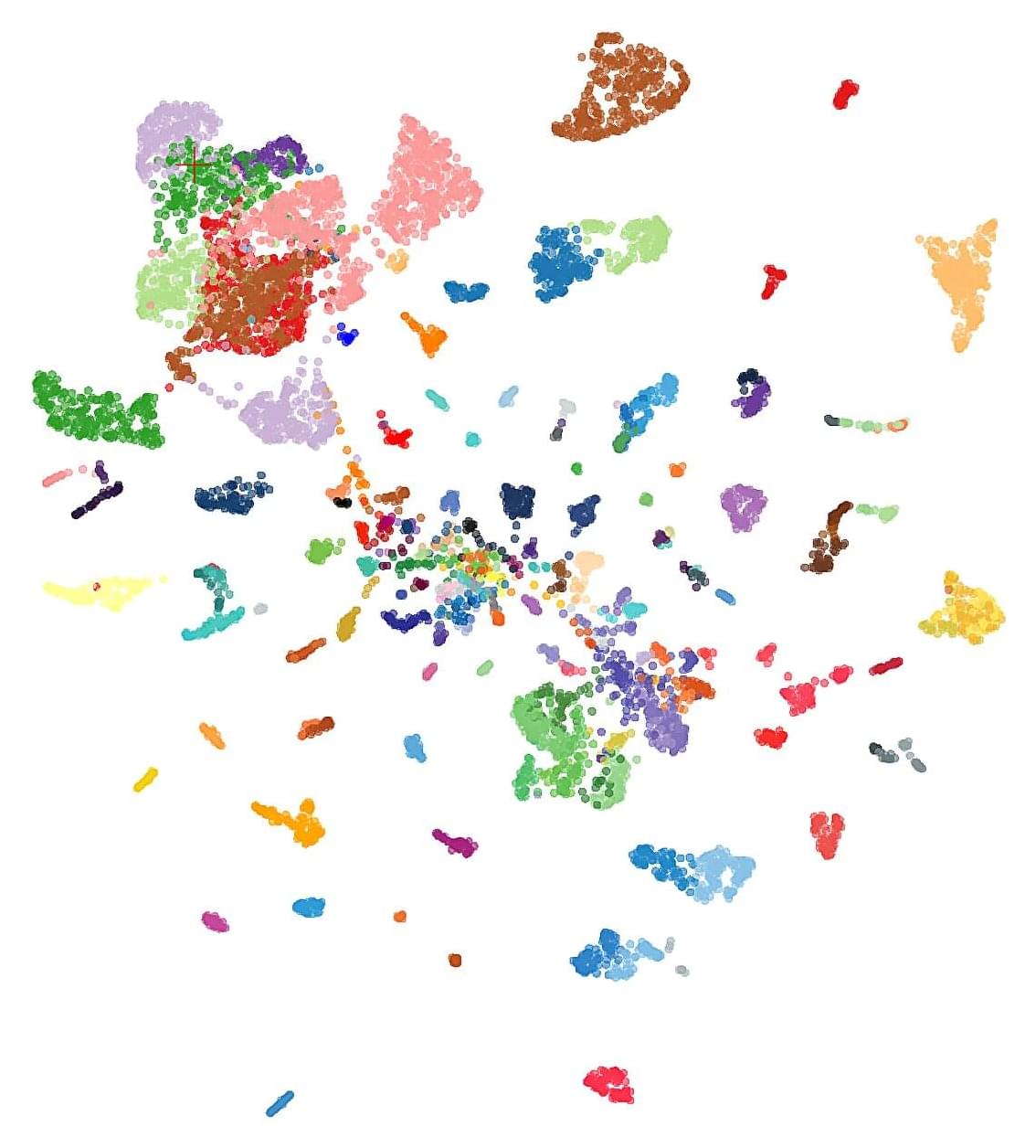
The model makes use of specific characteristics in the genetic material of tumors—their epigenetic fingerprint, obtained for example from , among other things. As the team shows in the journal Nature Cancer, the new model classifies tumors quickly and very reliably.

Today, far more types of tumors are known than the organs from which they arise. Each tumor has its own characteristics: certain tissue features, growth rates and metabolic peculiarities. Nevertheless, tumor types with similar molecular characteristics can be grouped together. The treatment of the individual disease depends decisively on the type of tumor.

YTHDFs as radiotherapy checkpoints in tumor immunity

N6-methyladenosine (m6A) is the most common and abundant endogenous mRNA methylation in eukaryotic cells (Huang et al., 2020; Wang et al., 2014). The regulation of this modification is achieved through the coordinated action of three distinct protein groups. The “writers” (methyltransferase complex), which include METTL3, METTL14, and WTAP, are responsible for adding the m6A modification. In contrast, the “erasers” (demethylases), which consist of FTO and ALKBH5, remove this chemical mark. Lastly, the “readers,” a group of proteins including YTHDF1/2/3 and YTHDC1/2, recognize and bind to m6A-modified RNA, thereby modulating diverse RNA metabolic processes. The m6A reader proteins YTHDFs exhibit distinct canonical functional roles: YTHDF1 primarily boosts the efficiency of mRNA translation, YTHDF2 enhances mRNA degradation, and YTHDF3 exerts dual functions by supporting both translation and degradation of mRNA, with its role varying depending on the specific biological context (Roundtree et al., 2017; Shi et al., 2017; Wang et al., 2014; Wang et al., 2015; Zaccara et al., 2019). Recent studies have revealed that YTHDF proteins can influence the efficacy of RT through mechanisms, such as modulating DNA repair and shaping the tumor immune microenvironment (TIME) (Du et al., 2023; Shao et al., 2023; Shi et al., 2023; Wang et al., 2023a; Wang et al., 2023c; Wen et al., 2024a; Yin et al., 2023). Elucidating the functions and mechanisms of YTHDF proteins within the context of radiation biology holds significant potential for advancing therapeutic strategies in cancer RT.

This review provides an overview of recent progress in elucidating the mechanisms by which YTHDF proteins in tumor and immune cells modulate the therapeutic efficacy of RT. By synthesizing current knowledge on the functions of YTHDF proteins in the context of IR, we emphasize their indispensable role in shaping RT outcomes.

/* */