From a vaccine for respiratory syncytial virus (RSV) to an infusion that slowed down Alzheimer’s for some people with the disease, here are three momentous advances from 2022.
An RSV vaccine showed promise for the first time in 50-years
Two vaccines are poised to be approved for RSV by the end of 2023, according to their makers, after almost 50-years without any meaningful progress.
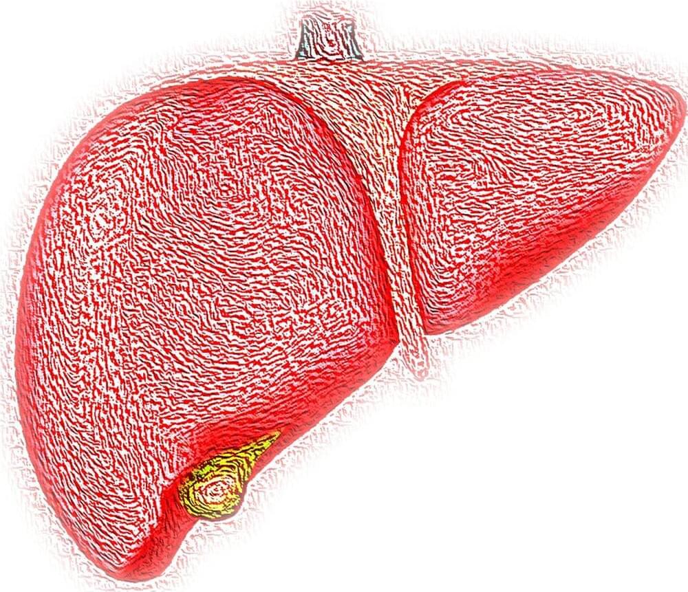
In a study examining the link between non-alcoholic fatty liver disease (NAFLD) and brain dysfunction, scientists at the Roger Williams Institute of Hepatology, affiliated to King’s College London and the University of Lausanne, found an accumulation of fat in the liver causes a decrease in oxygen to the brain and inflammation to brain tissue—both of which have been proven to lead to the onset of severe brain diseases.
The paper appears in the Journal of Hepatology.
NAFLD affects approximately 25% of the population and more than 80% of morbidly obese people. Several studies have reported the negative effects of an unhealthy diet and obesity can have on brain function however this is believed to be the first study that clearly links NAFLD with brain deterioration and identifies a potential therapeutic target.
Researchers in Spain have developed a new porous material capable of regenerating bones and preventing infections at the same time.
The scientists are from the Bioengineering and Biomaterials Laboratory of Universidad Católica de Valencia (UCV).
Tailor-made for each case using 3D printing, the biotech creations contain a bioactive alginate coating. This coating induces bone regeneration and destroys the bacteria that sometimes prevent bone formation from being completed.
Welcome to Futureunity, where we explore the fascinating world of science, technology, and the universe! From the inner workings of the human body to the outer reaches of space, we delve into the latest and most interesting discoveries that are shaping our world. Whether you’re a science buff or just looking for some mind-blowing facts, we’ve got you covered. Join us as we uncover the mysteries of the world around us and discover new frontiers in the fields of science and technology. Get ready for a journey that’s both educational and entertaining! Welcome to Futureunity, where we explore the fascinating world of science, technology, and the universe! From the inner workings of the human body to the outer reaches of space, we delve into the latest and most interesting discoveries that are shaping our world. Whether you’re a science buff or just looking for some mind-blowing facts, we’ve got you covered. Join us as we uncover the mysteries of the world around us and discover new frontiers in the fields of science and technology. Get ready for a journey that’s both educational and entertaining!
Disclaimer Fair Use: 1. The videos have no negative impact on the original works. 2. The videos we make are used for educational purposes. 3. The videos are transformative in nature. 4. We use only the audio component and tiny pieces of video footage, only if it’s necessary.
DISCLAIMER: Our channel is purely made for entertainment purposes, based on facts, rumors, and fiction.
Copyright Disclaimer under section 107 of the Copyright Act 1976, allowance is made for “fair use” for purposes such as criticism, comment, news reporting, teaching, scholarship, education, and research. Fair use is a use permitted by copyright statutes that might otherwise be infringing.
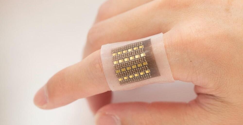
Electronic wearable patches have been devised to monitor various health conditions by noninvasively detecting biomolecules on the skin surface.
A new Nature Communicationsstudy discusses the development of novel skin patches capable of deep detection of biomolecules, which correlate better and more rapidly with physiological states. For example, the photoacoustic patch described by the researchers, who are engineers at the University of California San Diego, can produce a three-dimensional (3D) map of deep tissue hemoglobin.
Regenerative Medicine Daily is a news site dedicated to covering the latest breakthrough in the emerging field of regenerative medicine. We focus on scientific discoveries and research which hopes to allow medical science to exceed its current limitations.
T cells aren’t the first immune forces on the scene, they arrive after being alerted by other immune system warriors that a microbe has invaded or a cancer has silently seeded.
Exactly how T cells obtain the energy they need to build a massive army in the face of infiltrators has been the subject of speculation, theory and decades-long laboratory inquiries.
Now, scientists are taking a deeper dive into the question, and their investigations are shedding new light on an array of dynamic biological activities that help bolster T cell populations. Their research demystifies how T cells can power their growth and proliferation when disease emerges and T cell strength is in greatest need.