Toggle light / dark theme

Abstract: Detailing the effects of a new therapeutic tuberculosis vaccine!

https://doi.org/10.1172/JCI196648 In this Research Article, Styliani Karanika & team report on an intranasal DNA vaccine that accelerates TB cure and achieves better outcomes than standard or drug-resistant regimens alone in preclinical models.

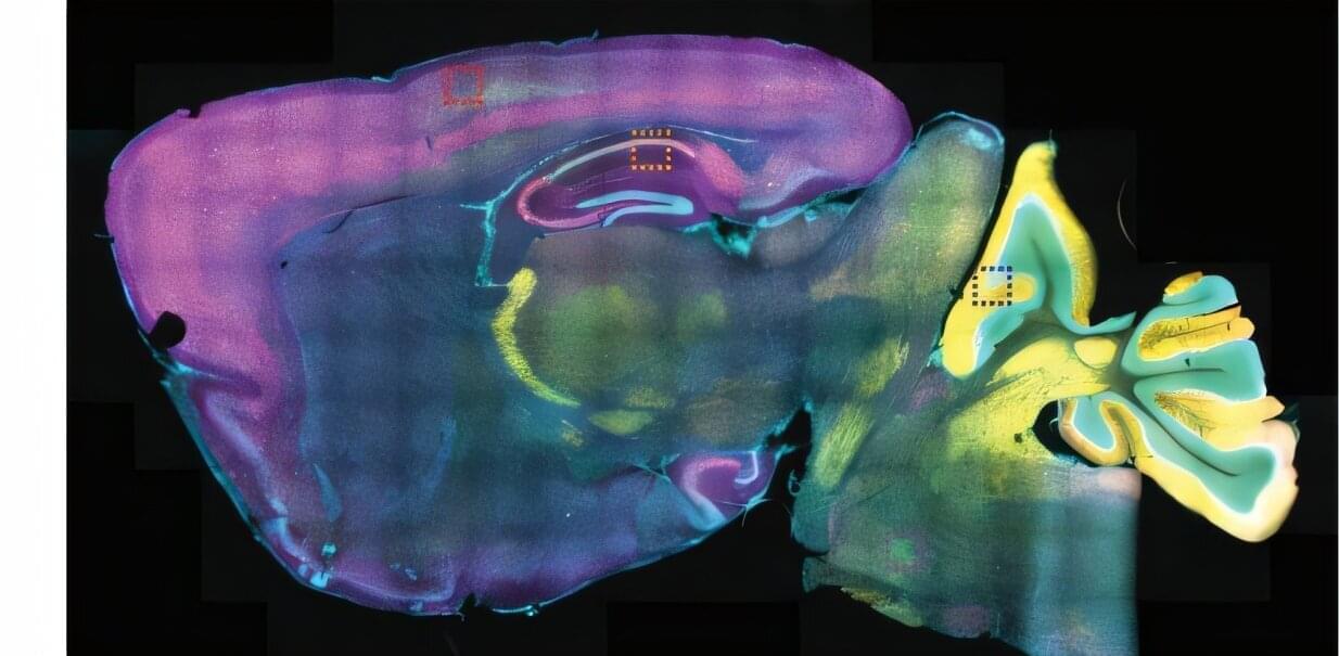
The figure shows mouse lungs with therapeutic intranasal Mip3a/relMt b fusion immunization, revealing local dendritic cell infiltration and enhanced colocalization with T cells.


1Center for Tuberculosis Research, Division of Infectious Diseases, Department of Medicine, Johns Hopkins University School of Medicine, Baltimore, Maryland, USA.

2W. Harry Feinstone Department of Molecular Microbiology and Immunology, Johns Hopkins Bloomberg School of Public Health, Baltimore, Maryland, USA.

3Division of Hematological Malignancies, Department of Oncology, Sidney Kimmel Comprehensive Cancer Center, Johns Hopkins University Hospital, Baltimore, Maryland, USA.

T-loop dynamics: telomere structure shapes cell fate decisions

Telomere structure shapes cell fate decisions.

Telomere loops (t-loops) are dynamic DNA structures, remodelled during the cell cycle and stress, rather than static protective caps.

The three-state model defines closed, intermediate, and uncapped telomeres, linking intermediate telomeres to programmed, fusion-resistant deprotection, which activates checkpoints without genome instability.

Mitotic arrest-dependent telomere deprotection is an active pathway in which Aurora B kinase drives t-loop unwinding without telomere shortening.

Aurora B kinase phosphorylation reprograms shelterin components (TRF1 and TRF2), enabling BTR-mediated t-loop dissolution and paradoxically converting protective factors into facilitators of deprotection.

T-loop dynamics reframe telomeres as responsive signalling hubs that couple chromosome architecture to genome surveillance and cell fate control. sciencenewshighlights ScienceMission https://sciencemission.com/T-loop-dynamics


Predicting Outcome After Newborn Stroke: A Lesion Network Mapping Study Leveraging Large-Scale Data

The development of cerebral palsy after neonatal stroke may be associated with disruptions of broad functional networks involving motor and extramotor regions as opposed to isolated lesions of the motor tracts.


Prior studies have established that resting-state networks are already present in newborns as early as term-equivalent age19 and that motor outcomes in healthy populations and other clinical populations are related to both primary motor networks and other broader motor networks.40,41 Our observation that the development of cerebral palsy correlated with both motor and extramotor regions lines up with these findings in other populations, and may indicate more widespread network vulnerabilities after stroke in neonates compared with adults.21 While it is known that disruption to primary motor regions is related to the development of cerebral palsy after NAIS,5,8 the extramotor regions (the frontal and temporal regions identified in the current study) may be more vulnerable to network-level disruptions in neonatal stroke due to the known relative immaturity of these regions during the neonatal period and their prolonged maturation to support higher-order functions.42,43 Particular vulnerability of these temporal and frontal regions has been demonstrated in other newborn, very preterm populations at risk of brain insult and injury.43

There are limitations to this study. Lesion network mapping has frequently been performed in adults and is more clinically feasible than directly acquiring resting-state fMRI scans in patients with NAIS; however, lesion network mapping may be seen as a less direct method for assessing brain connectivity.13,21,22 Functional connectivity has been well characterized in adults and neonates,1,16–19 but future studies will need to examine structural connectivity between regions via white matter fiber pathways using diffusion MRI scans to determine whether the functional changes identified correspond to structural changes.44 Such diffusion MRI investigations will be important in the future to investigate the roles (as part of global structural networks) of white matter regions and tracts such as the posterior limb of the internal capsule, which has previously been shown to be a strong predictor of motor outcome using individual region-based analyses.5–12 Given the paucity of prior research in this area, we aimed to establish that there is a relationship between the lesion functional connectome and cerebral palsy, but future work will also need to investigate the relative contributions of different measures (such as lesion volume, lesion location, and lesion functional and structural connectivity) to the development of cerebral palsy. Different measures may be more important for different brain regions (eg, lesions located in the primary motor regions are well-known to be related to cerebral palsy,5,8 whereas this study shows that functional connectivity of lesions to other subcortical and temporal and frontal cortical regions are related to cerebral palsy), meaning a combined approach considering multiple measures and regions may improve prediction of cerebral palsy in future work. Given that this study utilized clinically indicated MRIs from multiple sites for stroke participants, uniform scanner (eg, field strength) and sequence (eg, resolution) settings could not be used. The lesion network mapping approach does not necessarily require uniform scanning, making this approach more accessible. However, we acknowledge that variations in scanning parameters could influence some analysis steps, such as lesion segmentation and mapping to the template. To account for this, all analysis steps, including lesion segmentation and registration, were performed by experienced pediatric neurologists and image scientists, and outputs were extensively visually checked to ensure any poor-quality data were excluded.

This lesion network mapping approach identified correlations between lesions and the rest of the gray matter, which could include the equivalent lesion regions themselves (self-correlations, which are typically likely to be high). If self-correlations were very high in both groups (cerebral palsy and noncerebral palsy), then no significant group differences may be identified, which we think explains why the primary motor regions did not appear in the current findings (as the primary motor regions are key lesion regions affected by NAIS).5 The correlations identified between lesions and gray matter regions were positive (rather than negative correlations), which could reflect increased activity to support motor function (as opposed to decreased activity of irrelevant processes), as seen during task performance, but even intrinsically occurring at rest.45 Positive correlations are also, in general, stronger and less variable than negative correlations.46 We identified significant regions in both the left and right brain hemispheres; however, future work to specifically test whether there was a difference in the findings between hemispheres would be worthwhile (ie, to test whether results were greater in magnitude or spatial extent in the left or right hemisphere, which could be related to asymmetry in the lesions or connectome or both). With lesion network mapping, it was possible to infer that the regional networks identified correlated with cerebral palsy, but we cannot necessarily extrapolate on the relative importance of the individual regions of the network in cerebral palsy based on this method alone.13

Contribution of Life-Course Socioeconomic Position to Later-Life Brain Volumes in US Hispanic/Latino Adults

Among individuals with Schizophrenia and co-occurring SubstanceUseDisorders, psychological and psychosocial interventions showed limited benefit for symptom reduction and no effect on substance use, except for nicotine.


Question What is the efficacy of psychological and psychosocial interventions for individuals diagnosed with schizophrenia and comorbidity with substance use disorders?

Findings This systematic review and meta-analysis included 35 studies (4136 participants), with data available from 29 trials involving 3,831 participants covered in pairwise meta-analysis. Psychological and psychosocial interventions offered a very small effect on reducing patients’ overall symptoms, and no difference was found between intervention and control groups in reduction of all types of substance use intake or when separately analyzed, whereas nicotine use showed a modest improvement.

Meaning Current psychological and psychological interventions provide limited benefit for symptom reduction and were largely ineffective in decreasing substance use, except for a slight positive effect on nicotine use, indicating that more effective treatment strategies are urgently needed.

Cellular reprogramming beyond pluripotency

Aging, once viewed as an irreversible process, is now considered a modifiable process. Recent advances in cellular reprogramming reveal that transient expression of reprogramming factors can reverse molecular hallmarks of aging while preserving somatic cell identity. This ‘partial reprogramming’ rejuvenates tissues, restores regenerative capacity, and, in some models, extends lifespan without the tumorigenic risks of full dedifferentiation. In this review, we summarize genetic and chemical strategies for partial reprogramming, discuss their tissue-specific effects in vivo, and evaluate their implications for tissue regeneration and age-related disease. We further examine key challenges for clinical translation, including safety, delivery strategies, and temporal control of reprogramming.

Interstitial Lung Disease as a Late Occurrence in Ocrelizumab-Treated Patients With Multiple Sclerosis

Among patients with multiple sclerosis treated long-term with ocrelizumab, interstitial lung disease developed after a mean of 10.5 years, mainly as organizing pneumonia with variable outcomes.


This case series describes 6 cases of interstitial lung disease among patients with multiple sclerosis receiving long-term treatment with ocrelizumab.

Turmeric and ginger extract may boost implant bonding and kill 92% bacteria

An extract of turmeric and ginger helps bone implants bond strongly while killing bacteria and cancer cells, according to new research from Washington State University with implications for millions of patients with joint replacements and bone cancer. In early tests, the extract roughly doubled bone bonding within six weeks around the implant site, killed more than 90% of bacteria on implant surfaces, and sharply reduced cancer-causing cells. The findings marry elements of a naturopathic approach drawing on traditional medicine with current medical technologies. Turmeric, a golden-orange spice, and ginger root have been used for food and medicinal purposes in China and India for thousands of years.

“Basically, I say it’s combining the best with the latest,” said Susmita Bose, the Westinghouse Distinguished Chair Professor in WSU’s School of Mechanical and Materials Engineering and corresponding author of the paper. “The best part is from the food, and the latest aspect comes from the biomedical device.”

The new study, published in the Journal of the American Ceramic Society, is the most recent work from Bose and Amit Bandyopadhyay, Boeing Distinguished Professor in the School of Mechanical and Materials Engineering, demonstrating that compounds from turmeric and ginger can be effective supplements to cutting-edge medical treatment. That work builds upon their earlier research into the use of 3D printing to produce bone implants, an idea once considered far-fetched that is now a common way to manufacture implants.

3D microscopy reveals how a tick-borne virus reshapes human cells to replicate

Researchers at Umeå University show how tick-borne viruses remodel human cells into virus factories, using an advanced microscopy method. The findings provide new insight into how the virus replicates and matures, knowledge that may become important for future treatments against TBE. The study is published in Nature Communications.

“When we saw the three-dimensional images for the first time, we immediately realized how much new information we could gain about the virus’s replication,” says Lars-Anders Carlson, professor at the Department of Medical Chemistry and Biophysics at Umeå University, who led the study.

One of the most dangerous viral diseases spread in Europe is tick-borne encephalitis. A bite from an infected tick can transmit the TBE virus to humans and cause severe inflammation of the brain. Using electron microscopy, researchers at Umeå University have now discovered how tick-borne viruses reshape infected human cells and turn them into virus factories.

What this AI epitope library means for vaccines, immunotherapy and biosensors

A new tool makes it possible to screen millions of tiny protein fragments and select those that can be recognized by the immune system. The CIC biomaGUNE Center for Cooperative Research in Biomaterials has developed epiGPTope, a system that uses machine learning to generate and classify epitopes, in collaboration with the company Multiverse Computing.

The immune system is triggered by the presence of viruses or bacteria. When the antibodies produced recognize the epitopes, a small part of these viruses or bacteria, they launch an attack strategy. These epitopes are small fragments of protein recognized by antibodies or by immune cell receptors. So discovering new epitope sequences that target specific antibodies is essential for the development of diagnostic tools, immunotherapies and vaccines.

CIC biomaGUNE’s Biomolecular Nanotechnology laboratory, led by the Ikerbasque Research Professor Aitziber L. Cortajarena, is creating a library or database of hundreds of thousands of synthetic epitopes using this AI-based technique. The work is published in the journal ACS Synthetic Biology.

A layered approach sharpens brain signals in optical imaging

Near-infrared spectroscopy, or fNIRS, offers a way to monitor brain activity without surgery or radiation by tracking changes in blood flow and oxygenation. Light sources placed on the scalp send near-infrared light into the head, and detectors measure the light that scatters back. Because this light must pass through the scalp and skull before reaching the brain, the measured signal always includes a mix of superficial and cerebral contributions. Separating those signals has long been a central challenge for fNIRS researchers.

In a study published in Biophotonics Discovery, researchers from the Tufts University Diffuse Optical Imaging of Tissue Laboratory show that combining a specific source–detector geometry with a simple, anatomically informed tissue model can substantially improve how fNIRS data are interpreted.

By accounting for how light travels through layered head structures, the approach makes it possible to better isolate brain-specific signals without relying on complex imaging systems or subject-specific MRI scans.

/* */