A large project is underway to disease-proof pigs using CRISPR to change their DNA. Are people next?
Category: biotech/medical – Page 1529
Finding alternatives to antibiotics is one of the biggest challenges facing the research community. Bacteria are increasingly resistant to these drugs, and this resistance leads to the deaths of more than 25,000 around the world. Now, a multidisciplinary team of researchers from the Universitat Rovira i Virgili, the University of Grenoble (France), the University of Saarland (Germany) and RMIT University (Australia) have discovered that the mechanical deformation of bacteria is a toxic mechanism that can kill bacteria with gold nanoparticles. The results of this research have been published in the journal Advanced Materials and are a breakthrough in researchers’ understanding the antibacterial effects of nanoparticles and their efforts to find new materials with bactericide properties.
Since the times of Ancient Egypt, gold has been used in a range of medical applications and, more recently, as for diagnosing and treating diseases such as cancer. This is due to the fact that gold is a chemically inert material, that is, it does not react or change when it comes into contact with an organism. Amongst the scientific community, nanoparticles are known for their ability to make tumors visible and for their applications in nanomedicine.
This new research shows that these chemically inert nanoparticles can kill bacteria thanks to a physical mechanism that deforms the cell wall. To demonstrate this, the researchers have synthesized in the laboratory gold nanoparticles in the shape of an almost perfect sphere and others in the shape of stars, all measuring 100 nanometres (8 times thinner than a hair). The group analyzed how these particle interact with living bacteria. “We find that the bacteria become deformed and deflate like a ball that is having the air let out before dying in the presence of these nanoparticles,” explained Vladimir Baulin, researcher at the Department of Chemical Engineering of the URV. The researchers state the bacteria seem to have died after a massive leak, “as if the cell wall had spontaneously exploded.”
The mystery ailment that has afflicted U.S. embassy staff and CIA officers off and on over the last four years in Cuba, China, Russia and other countries appears to have been caused by high-power microwaves, according to a report released by the National Academies. A committee of 19 experts in medicine and other fields concluded that directed, pulsed radiofrequency energy is the “most plausible mechanism” to explain the illness, dubbed Havana syndrome.
The report doesn’t clear up who targeted the embassies or why they were targeted. But the technology behind the suspected weapons is well understood and dates back to the Cold War arms race between the U.S. and the Soviet Union. High-power microwave weapons are generally designed to disable electronic equipment. But as the Havana syndrome reports show, these pulses of energy can harm people, as well.
As an electrical and computer engineer who designs and builds sources of high-power microwaves, I have spent decades studying the physics of these sources, including work with the U.S. Department of Defense. Directed energy microwave weapons convert energy from a power source —a wall plug in a lab or the engine on a military vehicle—into radiated electromagnetic energy and focus it on a target. The directed high-power microwaves damage equipment, particularly electronics, without killing nearby people.
Dr. Carolina Reis Oliveria, is the CEO and Co-Founder of OneSkin Technologies, a biotechnology platform dedicated to exploring longevity science.
Carolina holds her Ph.D. in Immunology at the Federal University of Minas Gerais, in collaboration with the Rutgers University, where she conducted research with pluripotent stem cells as a source of retinal pigmented epithelium (RPE) cells, as well as the potential of RPE-stem cells derived as toxicological models for screening of new drugs with intra-ocular applications.
She founded a company called CELLSEQ solutions in Brazil which develops tools to revolutionize the safety and toxicology assays performed by pharmaceutical, cosmetic, agro-chemical and food industries, with technology based on stem cells and big data analysis.
She is an alumnus of IndieBio, the world’s leading biotechnology accelerator.
To visually illustrate the risk of airborne transmission in real time, The Washington Post used a military-grade infrared camera capable of detecting exhaled breath. Numerous experts — epidemiologists, virologists and engineers — supported the notion of using exhalation as a conservative proxy to show potential transmission risk in various settings.
“The images are very, very telling,” said Rajat Mittal, a professor of mechanical engineering in Johns Hopkins University’s medical and engineering schools and an expert on virus transmission. “Getting two people and actually visualizing what’s happening between them, that’s very invaluable.”
Doctors can triage and monitor patients faster—and sometimes more accurately—with the aid of the pocket-size machines.
Drexel University researchers are one step closer to offering a new treatment for the millions of patients who suffer from slow-healing, chronic wounds. The battery-powered applicator — as small and light as a watch — is the first portable and potentially wearable device to heal wounds with low-frequency ultrasound.
The National Institutes of Health (NIH) has awarded the research team an estimated $3 million to test the therapy on 120 patients over the next five years. By using diagnostic monitoring of blood flow in the wound tissue, the clinical trial will also determine how nutrition and inflammation impact wound closure, making treatment customization a possibility.
The project is an interdisciplinary collaboration between Drexel’s School of Biomedical Engineering, Science and Health Systems, the College of Medicine and the College of Nursing and Health Professions.
Circa 2008
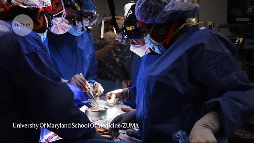
Three independent research teams have successfully performed organ transplantations that do not require the recipient to face a lifetime of immunosuppressant drugs to prevent rejection. Instead, the new techniques prevent rejection by training the immune system to recognize the new organ as its own.
The three studies, published this week in the New England Journal of Medicine, are preliminary and involve only a few patients. But if the techniques can be reproduced in a larger population, they could eliminate one of the most enduring scars of the operation: the need to continue taking sometimes-dangerous immunosuppressant drugs.
Thousands of kidney transplantations are performed every year, and nearly 99% of patients in the United States are still alive a year after the surgery. But even when the organ donor is a close relative, the transplant recipient often needs to take immunosuppressant drugs for the rest of their lives to guard against organ rejection. But although the drugs help to prevent rejection, they also increase the risk of infection and are very pricey.
The organ-on-a-chip (OOAC) is in the list of top 10 emerging technologies and refers to a physiological organ biomimetic system built on a microfluidic chip. Through a combination of cell biology, engineering, and biomaterial technology, the microenvironment of the chip simulates that of the organ in terms of tissue interfaces and mechanical stimulation. This reflects the structural and functional characteristics of human tissue and can predict response to an array of stimuli including drug responses and environmental effects. OOAC has broad applications in precision medicine and biological defense strategies. Here, we introduce the concepts of OOAC and review its application to the construction of physiological models, drug development, and toxicology from the perspective of different organs. We further discuss existing challenges and provide future perspectives for its application.
Brain on a chip for drug discovery.
Since the advent of organ-on-a-chip, many researchers have tried to mimic the physiology of human tissue on an engineered platform. In the case of brain tissue, structural connections and cell–cell interactions are important factors for brain function. The recent development of brain-on-a-chip is an effort to mimic those structural and functional aspects of brain tissue within a miniaturized engineered platform. From this perspective, we provide an overview of trace of brain-on-a-chip development, especially in terms of complexity and high-content/high-throughput screening capabilities, and future perspectives on more in vivo-like brain-on-a-chip development.
With the advent of an aging society, the disease incidence rate is increasing, and the cost of drug development and disease treatment is expanding exponentially.1,2 According to the World Health Organization (WHO), nearly one billion people in the world suffer from neurodegenerative diseases such as Alzheimer’s (AD) and Parkinson’s diseases.3 Despite decades of research on neurodegenerative diseases by many biologists and pharmaceutical companies, the underlying mechanism of their onset and progression is still largely unknown. The resolution of these diseases has a long way to go, and such steps are limited due to the lack of a suitable in vitro model system for mechanism study and drug development. In particular, the complex tissue structures and cell–cell interactions of the in vivo system make it challenging to unravel the underlying mechanism of the diseases and to predict the efficacy of clinical medicine.