From marigolds to human babies, most complex organisms start as a single-celled embryo. In his new book, Ben Stanger explores what our humble origins could teach us about health and disease.
By Clare Wilson

From marigolds to human babies, most complex organisms start as a single-celled embryo. In his new book, Ben Stanger explores what our humble origins could teach us about health and disease.
By Clare Wilson
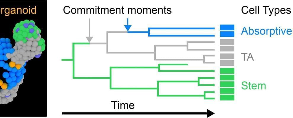
AMOLF researchers discovered that stem cells first specialize into a functional cell and then move to their proper location—rather than the other way around.
Researchers at AMOLF, Amsterdam, and the Hubrecht institute, Utrecht, revealed a new model to show how stem cells specialize into functional cells. They found that their position in the organ is not as important as current models claim. Rather, stem cells choose their identity first and only then move to their appropriate position.
These discoveries were made using intestinal organoids and the new TypeTracker technique, which can now be used to understand other organs at the cellular level and the effects of mutations and medications. The findings were published on August 18 in the journal Science Advances.

The research, published in Proceedings of the National Academy of Sciences, used a genetic approach to fix deafness in mice with a defective Spns2 gene, restoring their hearing abilities in low and middle frequency ranges. Researchers say this proof-of-concept study suggests that hearing impairment resulting from reduced gene activity may be reversible.
Over half of adults in their 70s experience significant hearing loss. Impaired hearing is associated with an increased likelihood of experiencing depression and cognitive decline, as well as being a major predictor of dementia. While hearing aids and cochlear implants may be useful, they do not restore normal hearing function, and neither do they halt disease progression in the ear. There is a significant unmet need for medical approaches that slow down or reverse hearing loss.
New research from the Institute of Psychiatry, Psychology & Neuroscience (IoPPN) at King’s College London has successfully reversed hearing loss in mice.
This proof-of-concept study suggests that gene therapy for this type of hearing loss in humans may be successful in the future.

I will try to live as long as possible.
Dr. Ezekiel Emanuel plans to reject life-extending medical care at the age of 75. The reason he does this is quite similar to why the Kaelons commit ritual suicide in Star Trek: The Next Generation. Does this make sense?
In this thought-provoking episode of Lifespan News, host Ryan O’Shea delves deep into the controversial topic of choosing when to die and the ethics surrounding medical interventions to prolong life. Using the lens of a Star Trek: The Next Generation episode and drawing parallels with Dr. Ezekiel Emanuel’s The Atlantic article, “Why I Hope to Die at 75″, Ryan confronts the moral and societal implications of setting an arbitrary age to stop seeking medical treatment. With advancements in rejuvenation biotechnologies, is it reasonable to maintain such views? As we push the boundaries of science and healthcare, when should we draw the line? Join Ryan as he navigates these complex questions, and remember to share your thoughts in the comments below. Don’t forget to subscribe for more!
Video Clips:
Leading US doctor says he won’t get treatment if he gets cancer after 75, CNN — https://www.youtube.com/watch?v=TgrO4rrrFgQ
How Long Do You Want to Live?, The Atlantic — https://youtu.be/fQBzY-aorFQ

The FDA on Friday approved Regeneron Pharmaceuticals’ Veopoz (pozelimab-bbfg), the first and only treatment indicated specifically for CHAPLE disease, also known as CD55-deficient protein-losing enteropathy, according to the company.
A fully human monoclonal antibody, Veopoz is approved for the treatment of adult and pediatric patients 1 year of age and older with CHAPLE, an ultra-rare hereditary disease that can cause potentially life-threatening gastrointestinal and cardiovascular symptoms.
- CHAPLE—which stands for complement hyperactivation, angiopathic thrombosis, and protein-losing enteropathy—is an inherited immune disease that causes the complement system (the part of your immune system that defends the body against injury and foreign invaders like bacteria and viruses) to become overactive.-FDA.
The regulator’s greenlight on Friday for Regeneron Pharmaceuticals’ monoclonal antibody Veopoz (pozelimab-bbfg) makes it the first and only treatment indicated for children and adults with CHAPLE disease.

Contact Lens Health Week will be observed Aug. 21–25, which makes this a good time to learn more about contact lenses and whether they might be right for you.
Eyeglasses can be fun and fashionable. And they’re a safe and effective way to provide vision correction for most people. Contact lenses also can provide a safe and effective way to correct your vision, and more than 45 million people in the U.S. wear contact lenses, according to the Centers for Disease Control and Prevention.
If you’re considering switching to contact lenses, here are some things you should consider.

Hundreds of elders are using an AI-powered robot to combat their loneliness — and it appears to be offering companionship to one New Yorker getting daily breast cancer treatments.
Priscilla O’Kesson, a 77-year-old, told Spectrum News she has been using ElliQ, an AI-powered robot made by Israeli startup Intuition Robotics, to keep her company at her home in a town in Greene County called Catskill.
The AI-robot, designed to help older adults age on their own, uses voice commands and on-screen instructions to talk to and interact with users in real-time. ElliQ can conduct daily check-ins, act as a personal trainer, play games to stimulate the brain, and track their wellness goals, according to the robotics company.
Group A streptococci are one of the most common pathogens that humans are exposed to, and they can cause infections with a wide range of severities, from mild rashes and sore throats to flesh-eating and systemic infections that can be fatal. The number of these infections is also on the rise, although the reasons are unclear. Now researchers have learned more about why these pathogens can be mild in some people, and hit others hard. The findings have been reported in Nature Communications.
Scientists suspected that some interaction between a person’s genetics and the bacterial pathogen could be leading to such varied outcomes, said study co-author Fredric Carlsson, a researcher at Lund University.
The National Institute of Allergy and Infectious Diseases (NIAID), one of the largest institutes in the National Institutes of Health (NIH), part of the Department of Health and Human Services (HHS), conducts and supports basic and applied research to better understand, treat, and ultimately prevent infectious, immunologic, and allergic diseases. The Bacterial Pathogenesis and Antimicrobial Resistance Unit (chief John Dekker) in the Laboratory of Clinical Immunology and Microbiology in the Division of Intramural Research (DIR) within NIAID seeks candidates for a postdoctoral fellowship position in microbial genomics.
This position will offer a unique opportunity to work at the intersection of pathogen genomics, systems biology, and clinical infectious diseases. The lab uses a variety of genomics, transcriptomics, metabolomics, imaging, and molecular approaches to study bacterial pathogens and antimicrobial resistance, with a focus on intra-host evolution in the context of infection. Access to state-of-the-art short-and long-read sequencing, metabolomics, optical, and computational resources is available. See more information about the Bacterial Pathogenesis and Antimicrobial Resistance Unit under chief John P. Dekker and an example of their recent work.