The Utah-based pharmaceutical company has trained a large language model to enable scientists to tap into dozens of machine learning models at once, saving them time during drug development.

Year 2021 face_with_colon_three
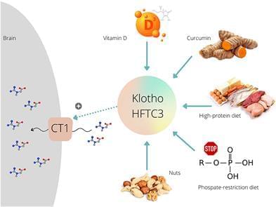
Creatine plays a pivotal role in cellular bioenergetics, acting as a temporal and spatial energy buffer in cells with high and fluctuating energy requirements (1). Jeopardizing delicate creatine homeostasis can be detrimental to many energy-demanding tissues, including the brain. For instance, cerebral creatine hypometabolism accompanies various neurological conditions, including a number of developmental disorders (2, 3), neurodegenerative and cerebrovascular diseases (4, 5), and brain cancer (6). A reduced creatine availability in the brain has been thus recognized as an apposite therapeutic target, and supplying exogenous creatine to compensate for a disease-driven shortfall emerged as a first possible approach. However, early success in animal models of neurological diseases was not corroborated in human trials, with the use of creatine supplementation proved largely disappointing in clinical studies with a number of symptomatic neurological disorders [for a detailed review, see (7)]. A meager delivery of creatine to the brain could be partly due to a low activity/density of creatine transporter (CT1 or SLC6A8), a transmembrane sodium-and chloride-dependent protein that mediates creatine uptake into the target cells (8). For that reason, the upregulation of CT1 function has been identified as an innovative course of action to facilitate creatine uptake, with several exotic agents and routes were cataloged so far, including glucocorticoid-regulated kinases, mammalian target of rapamycin, ammonia, and Klotho protein (9).
Besides other vehicles, Klotho protein (Clotho; HFTC3) is put forward as a possible stimulator of CT1 function that can uplift creatine allocation to the target tissues. This membrane-bound pleiotropic enzyme (also exists in a circulating form) participates in many metabolic pathways, including calcium-phosphate metabolism, nutrient sensing, and remyelination (10). Klotho is highly expressed in neuronal cells of the cerebral cortex, cerebellum, and spinal cord (11). The role of Klotho in high-phosphate energy metabolism modulation was revealed a few years ago when Amilaji et al. (12) found that the co-expression of Klotho protein increases a creatine-induced current in CT1-expressing cells. The authors reported that the current through CT1 was a function of the extracellular creatine levels, with the maximal creatine-induced current was higher in cells expressing CT1 together with Klotho than in cells expressing CT1 alone (29.5 vs. 20.2 nA).
Laboratory “copilots” and automated labs are AI’s latest contribution to speeding up the development of new drugs, chemicals and materials. Why it matters: Scientific discovery itself must speed up if the world is to address its challenges — from climate change to personalized treatments for cancer — fast enough to make a difference. In scientific research, “manual effort is not scalable,” writes Microsoft Health Futures’ Hoifung Poon in the…
The first functional semiconductor made from graphene has been created at the Georgia Institute of Technology. This could enable smaller and faster electronic devices and may have applications for quantum computing.
Credit: Georgia Institute of Technology.
Semiconductors, which are materials that conduct electricity under specific conditions, are foundational components of electronic devices like the chips in your computer, laptop, and smartphone. For many decades, their architecture has been getting smaller and more compact – a trend known as Moore’s Law. This has enabled gigantic leaps in a vast range of technologies, from general computing speeds and video game graphics, to the resolution of medical scans and the sensitivity of astronomical observatories.
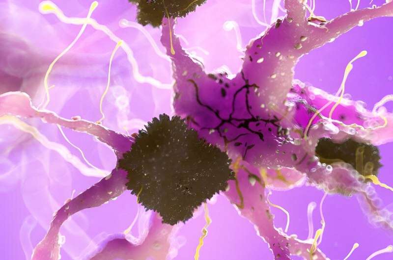
The therapy—developed at the University of Nebraska Medical Center (UNMC)—relies on both the immune system to fight key aspects of Alzheimer’s, plus modified cells that zero in on the brain protein plaques that are a hallmark of the disease.
In patients with Alzheimer’s, amyloid-beta protein forms plaques that prevent nerve cells from signaling each other. One theory is that this might cause irreversible memory loss and behavior changes characteristic of Alzheimer’s disease.
The new study was recently published in the journal Molecular Neurodegeneration. Researchers used genetically modified immune-controlling cells called Tregs to target amyloid-beta.
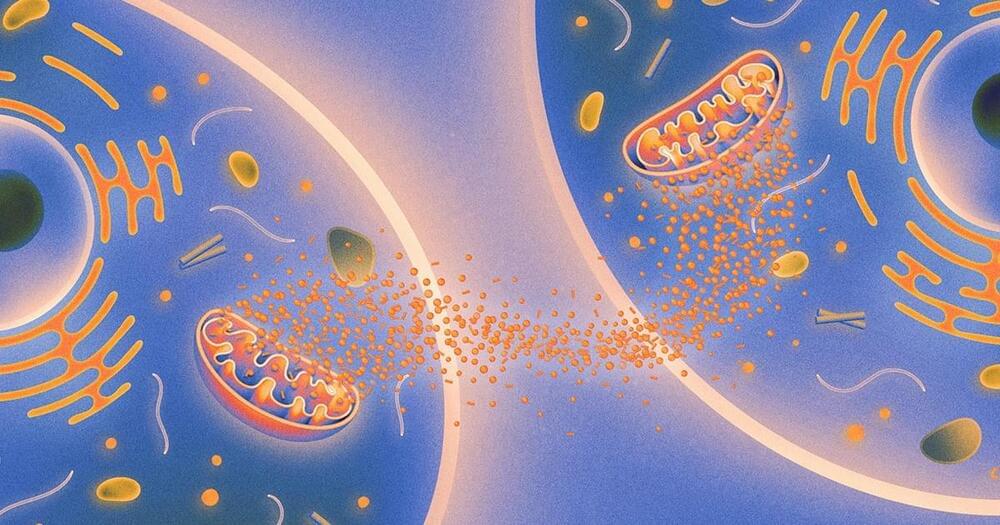
The research builds on a recent body of work that suggests that #mitochondria are social organelles that can talk to one another even when they are in different tissues.
Biologists discovered that mitochondria in different tissues talk to each other to repair injured cells. When their signal fails, the biological clock starts winding down.
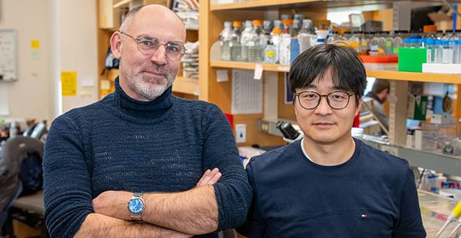
A new study published in Nature Cell Biology by Mark Alkema, PhD, professor of neurobiology, establishes an important molecular link between specific B12-producing bacteria in the gut of the roundworm C. elegans and the production of acetylcholine, a neurotransmitter important to memory and cognitive function.
There is growing recognition among scientists that diet and gut microbiota may play an important role in brain health. Changes in the composition of the microbiome have been linked to neurological disorders such as anxiety, depression, migraines and neurodegeneration. Yet, teasing out the cause and effect of individual bacteria or nutrients on brain function has been challenging.
“There are more bacteria in your intestine than you have cells in your body,” said Woo Kyu Kang, PhD, a postdoctoral fellow in the Alkema lab and first author of the current study. “The complexity of the brain, the hundreds of bacterial species that comprise the gut microbiome and the diversity of metabolites make it almost impossible to discern how bacteria impact brain function.”

“Epigenetic changes determine whether genes are turned on or off, and can potentially reverse disease, broadening the therapeutic landscape to find potential cures previously thought impossible.”
Company co-founded by Alex Aravanis and Feng Zhang targets epigenetic code to reprogram cells to a healthy state.
