“If you look at the brain chemically, it’s like a soup with a bunch of ingredients,” said Dr. Fan Lam.
Can we map the brain to show its behavior patterns when a patient is healthy and sick? This is what a recent study published in Nature Methods hopes to address as a team of researchers at the University of Illinois Urbana-Champaign used a $3 million grant obtained from the National Institute of Aging to develop a novel approach to mapping brain behavior when a patient is both healthy and sick. This study holds the potential to help researchers, medical professionals, and patients better understand how to treat diseases.
“If you look at the brain chemically, it’s like a soup with a bunch of ingredients,” said Dr. Fan Lam, who is an assistant professor of bioengineering at the University of Illinois Urbana-Champaign and a co-author on the study. “Understanding the biochemistry of the brain, how it organizes spatiotemporally, and how those chemical reactions support computing is critical to having a better idea of how the brain functions in health as well as during disease.”
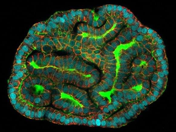
For the study, the researchers used a type of technology called spatial omics and combined this with deep learning to produce 3D datasets to unveil the brain’s myriad of characteristics down to the molecular level. Through this, the team has developed a novel method in monitoring brain activity when a patient is both healthy and sick, including the ability to identify complex neurological diseases.


