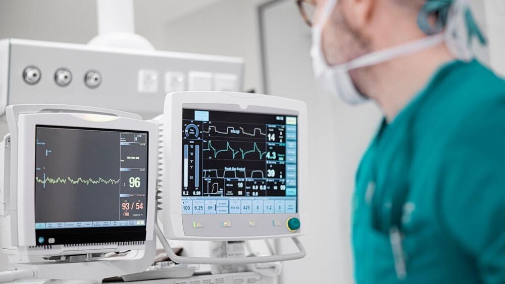
UC Riverside unveils real-time visibility and 10x sensitivity. These could revolutionize chronic back pain treatment.

Organic computers are based on living, biological “wetware”. This video reports on organic computing research in areas including DNA storage and massively parallel DNA processing, as well as the potential development of biochips and entire biocomputers. If you are interested in this topic you may enjoy my book “Digital Genesis: The Future of Computing, Robots and AI”. You can download a free pdf sampler, here: http://www.explainingcomputers.com/ge… purchase “Digital Genesis” on Amazon.com here: http://amzn.to/2yVKStK Or purchase “Digital Genesis” on Amazon.co.uk here: http://www.amazon.co.uk/dp/1976098068… Links to specific research cited in the video are as follows: Professor William Ditto’s “Leech-ulator”: http://www.zdnet.com/article/us-scien… Development of transcriptor at Stanford: https://med.stanford.edu/news/all-new… Harvard Medical School DNA storage: https://hms.harvard.edu/news/writing–… Yaniv Erlich and Dina Zielinski DNA storage: http://pages.jh.edu/pfleming/bioinfor… Manchester University DNA parallel processing: http://rsif.royalsocietypublishing.or… All biocomputer and other CG animations included in this video were produced by and are copyright © Christopher Barnatt 2017. If you enjoy this video, you may like my previous report on quantum computing: • Quantum Computing 2017 Update More videos on computing-related topics can be found at:
/ explainingcomputers You may also like my ExplainingTheFuture channel at:
/ explainingthefuture.
For the survival of life on Earth, the process where plants perform photosynthesis to generate oxygen and chemical energy using sunlight is crucial. Scientists from Göttingen and Hannover have now achieved a breakthrough by creating a high-resolution 3D visualization of the chloroplasts’ copying mechanism, the RNA polymerase PEP, for the first time. This intricate structure offers fresh perspectives on the operation and evolutionary history of this vital cellular apparatus, instrumental in interpreting the genetic blueprints for proteins involved in photosynthesis.
Without photosynthesis, there would be no air to breathe – it is the basis of all life on Earth. This complex process allows plants to convert carbon dioxide and water into chemical energy and oxygen using light energy from the sun. The conversion takes place in the chloroplasts, the heart of photosynthesis. Chloroplasts developed in the course of evolution when the ancestors of today’s plant cells absorbed a photosynthetic cyanobacterium. Over time, the bacterium became increasingly dependent on its “host cell”, but maintained some significant functions such as photosynthesis and parts of the bacterial genome. The chloroplast therefore still has its own DNA, which contains the blueprints for crucial proteins of the “photosynthesis machinery”
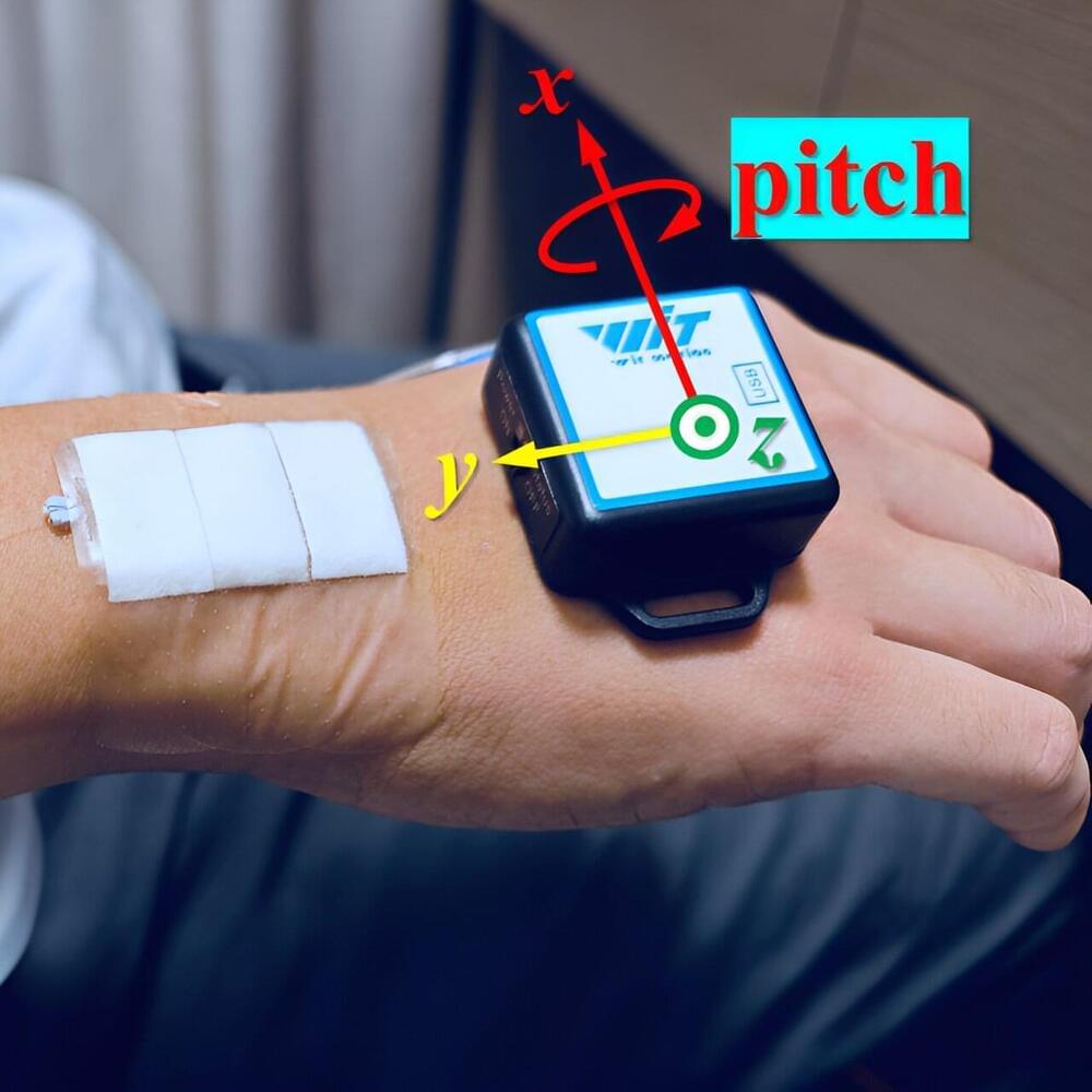
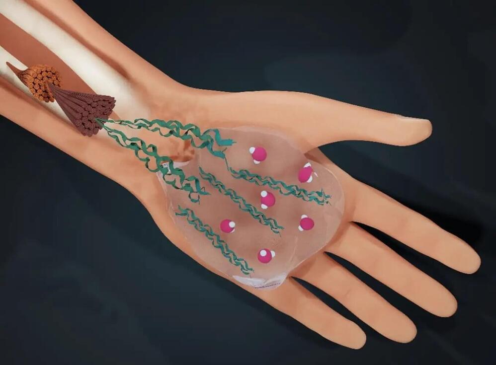
Imagine wearing a thin flexible sticker that can turn your hand or finger movement into communication without you having to say a word or tap a touch screen. Researchers have developed a new type of wearable sensor that can accomplish this futuristic feat and could open new possibilities for rehabilitation applications and help those with disabilities to communicate more easily.
The new sensor combines a soft and flexible material called polydimethylsiloxane, or PDMS, with an optical component known as a fiber Bragg grating (FBG). The researchers designed it to be comfortable for long-term wear while also having the ability to detect movements with high accuracy.
A paper describing this technology is published in the journal Biomedical Optics Express.
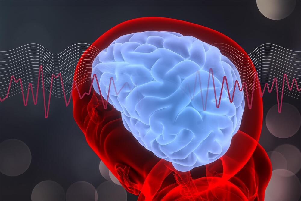
Stimulating gamma brain waves may protect cancer patients from memory impairment and other cognitive effects of chemotherapy.
Patients undergoing chemotherapy often experience cognitive effects such as memory impairment and difficulty concentrating — a condition commonly known as “chemo brain.”
MIT…
A noninvasive treatment may help to counter “chemo brain” impairment often seen in chemotherapy patients: Exposure to light and sound with a frequency of 40 hertz protected brain cells from chemotherapy-induced damage in mice, MIT researchers found.
Recent research challenges the long-standing understanding of the end-replication problem in DNA, revealing two distinct issues rather than one.
Half a century ago, scientists Jim Watson and Alexey Olovnikov independently realized that there was a problem with how our DNA gets copied. A quirk of linear DNA replication dictated that telomeres that protect the ends of chromosomes should have been growing shorter with each round of replication, a phenomenon known as the end-replication problem.
Telomerase: A Solution Emerges
Water determines life: humans are three-quarters water. An international research team led by the University of Amsterdam (UvA) has now discovered how water also determines the structure of the material that holds us together: collagen.
In a paper published in PNAS, the researchers elucidate the role of water in the molecular self-assembly of collagen. They show that by replacing water with its ‘twin molecule’ heavy water (D2O), one can ‘tune’ the interaction between collagen molecules, and thus influence the process of collagen self-assembly. The findings will help to better understand the tissue failures resulting from heritable collagen-related diseases, such as brittle bone disease (osteogenesis imperfecta).
As lead author Dr. Giulia Giubertoni of the UvA’s Van ‘t Hoff Institute for Molecular Sciences (HIMS) puts it, “In studying these and other collagen diseases, many researchers, including myself, … have always missed an important part of the puzzle, and the possibility that tissue failure might be partly due to water-collagen interaction was not taken very seriously. We now show that perturbing the water layer around the protein, even very slightly, has dramatic effects on collagen assembly.”