Using a new technique, scientists created genetic blueprints for kangaroos, penguins, sharks and more.


The need to quash outbreaks, quickly create medicines, stress-proof crops and fend off other 21st century threats is providing a lucrative arena for biotech companies to sell their services.
Why it matters: But the infrastructure to support such ambitions is increasingly recognized by the U.S., China and other countries as a linchpin of national security and economic strategy, putting it at the center of geopolitics.
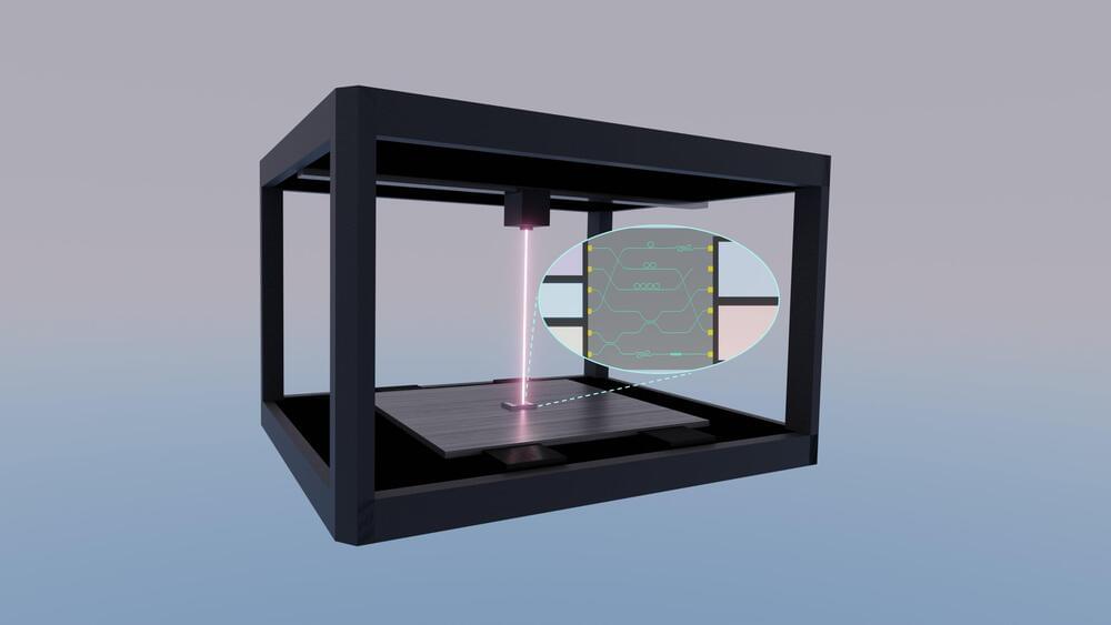
Photonic integrated circuits are an important next-wave technology. These sophisticated microchips hold the potential to substantially decrease costs and increase speed and efficiency for electronic devices across a wide range of application areas, including automotive technology, communications, health care, data storage, and computing for artificial intelligence.
Photonic circuits use photons, fundamental particles of light, to move, store, and access information in much the same way that conventional electronic circuits use electrons for this purpose. Photonic chips are already in use today in advanced fiber-optic communication systems, and they are being developed for implementation in a broad spectrum of near-future technologies, including light detection and ranging, or LiDAR, for autonomous vehicles; light-based sensors for medical devices; 5G and 6G communication networks; and optical and quantum computing.
Given the broad range of existing and future uses for photonic integrated circuits, access to equipment that can fabricate chip designs for study, research and industrial applications is also important. However, today’s nanofabrication facilities cost millions of dollars to construct and are well beyond the reach of many colleges, universities, and research labs.

The lack of representation of Asians in the genome can cause “large deviations” when diagnosing or treating patients, and could affect the development of targeted drugs, he said.
To address the gap, in 2020 Gao and his research team set out to construct a reference of the Chinese genome, particularly of the Han ethnicity, the largest ethnic group in the world.
Reverse Aging Revolution
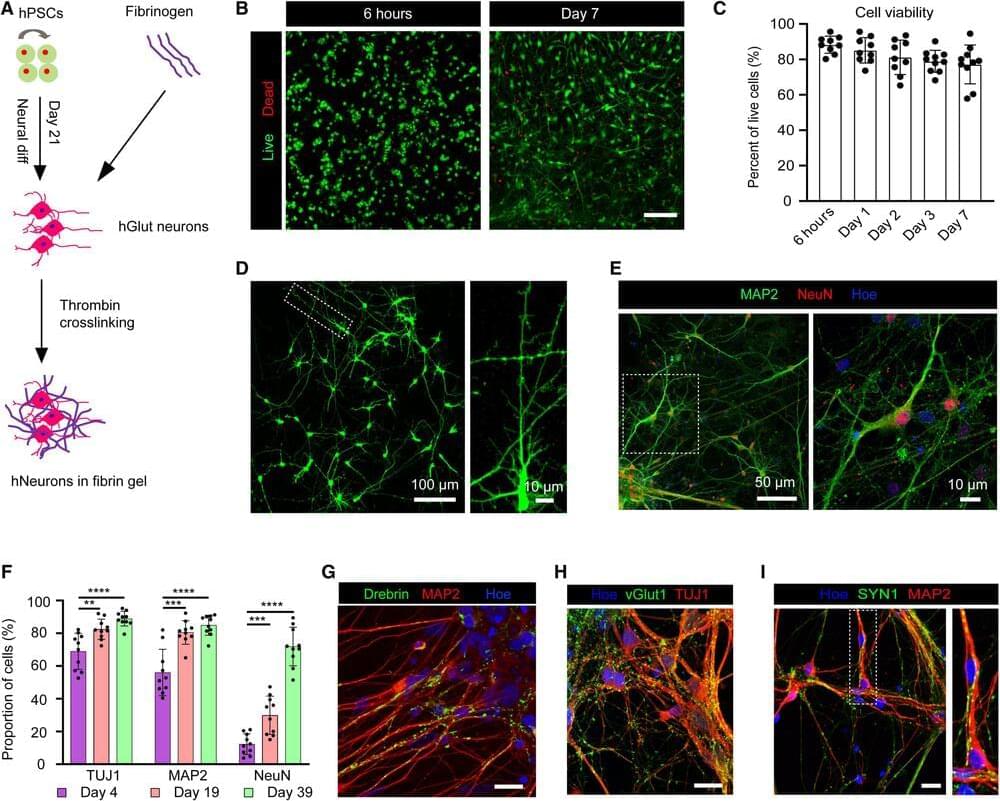
A team of University of Wisconsin–Madison scientists has developed the first 3D-printed brain tissue that can grow and function like typical brain tissue.
It’s an achievement with important implications for scientists studying the brain and working on treatments for a broad range of neurological and neurodevelopmental disorders, such as Alzheimer’s and Parkinson’s disease.
“This could be a hugely powerful model to help us understand how brain cells and parts of the brain communicate in humans,” says Su-Chun Zhang, professor of neuroscience and neurology at UW–Madison’s Waisman Center. “It could change the way we look at stem cell biology, neuroscience, and the pathogenesis of many neurological and psychiatric disorders.”
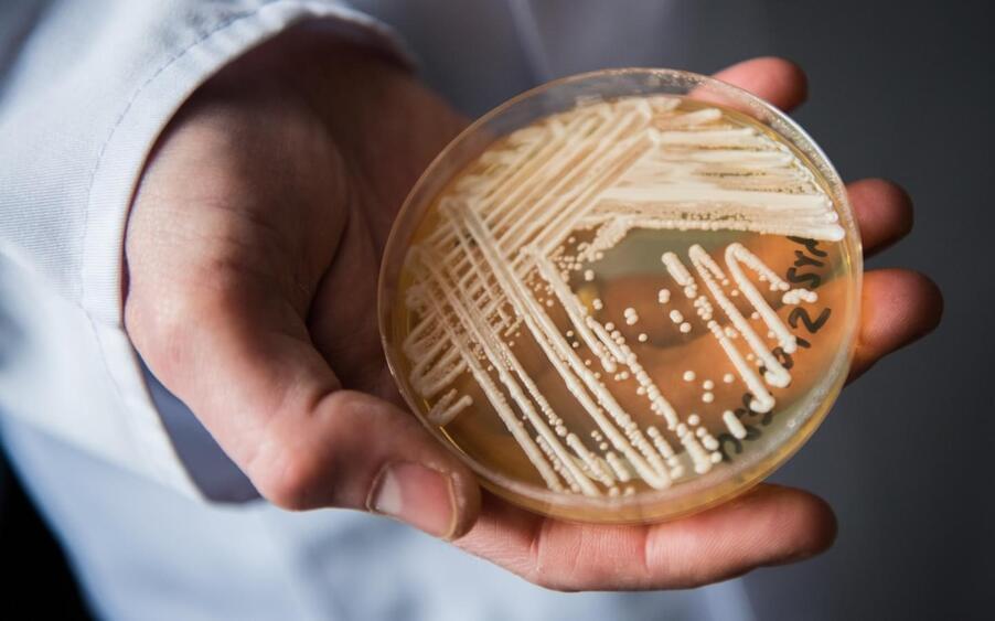
Washington state is experiencing its first known outbreak of a potentially deadly fungus, according to public health officials.
Four patients in the last month have tested positive for Candida auris, or C. auris, Public Health — Seattle & King County said in a release.
The first case occurred in a patient who had recently been admitted to Kindred Hospital Seattle, which was identified through a proactive screening program.

The rare condition posterior cortical atrophy (PCA) involves strange, troubling issues with vision and spatial awareness – including difficulty judging distances, seeing movement, and recognizing objects – and a new study highlights its close relationship to Alzheimer’s disease in more detail than ever before.
PCA and Alzheimer’s have long been linked with each other, because they share a lot of the same pathological changes in the brain. However, the rarity of PCA has made it hard for researchers to fully assess it in relation to Alzheimer’s.
To address that, an international team of researchers analyzed data on 1,092 individuals with PCA, finding that it was a very strong predictor for Alzheimer’s: in 94 percent of cases, tell-tale Alzheimer’s brain changes were observed, and were most likely contributing to PCA.
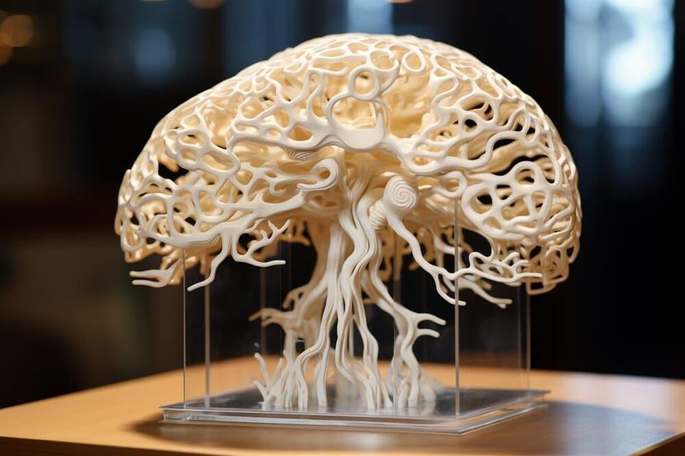
Summary: Researchers developed the world’s first 3D-printed brain tissue that grows and behaves similarly to natural brain tissue, marking a significant leap forward for neurological and neurodevelopmental disorder research.
This novel 3D-printing technique uses a horizontal layering approach and a softer bio-ink, allowing neurons to interconnect and form networks akin to human brain structures.
The ability to precisely control cell types and arrangements provides unparalleled opportunities to study brain functions and disorders in a controlled environment, offering new avenues for drug testing and understanding brain development and diseases like Alzheimer’s and Parkinson’s.