Toggle light / dark theme

In recent years, ribonucleic acid (RNA) has emerged as a powerful tool for the development of novel therapies. RNA is used to copy genetic information contained in our hereditary material, the deoxyribonucleic acid (DNA), and then serves as a template for building proteins, the building blocks of life. Delivery of RNA into cells remains a major challenge for the development of novel therapies across a broad range of diseases. Researchers at the Max Planck Institute of Molecular Cell Biology and Genetics (MPI-CBG) in Dresden together with researchers from the global biopharmaceutical company AstraZeneca have investigated where and how mRNA is delivered inside the cell. They found that mRNA uses an unexpected entry door. Their results provide novel insights into the development of RNA therapeutics towards efficient delivery and lower dosages.

DNA () contains the required for the development and maintenance of life. This information is communicated by messenger (mRNA) to make proteins. mRNA-based therapeutics have the potential to address unmet needs for a wide variety of diseases, including cancer and cardiovascular disease. mRNA can be delivered to cells to trigger the production, degradation or modification of a target protein, something impossible with other approaches. A key challenge with this modality is being able to deliver the mRNA inside the cell so that it can be translated to make a protein. mRNA can be packed into lipid nanoparticles (LNPs)—small bubbles of fat—that protect the mRNA and shuttle it into cells. However, this process is not simple, because the mRNA has to pass the membrane before it can reach its site of action in the cell interior, the cytoplasm.

Researchers in the team of MPI-CBG director Marino Zerial are experts in visualizing the cellular entry routes of molecules in the cell, such as mRNA with high-resolution microscopes. They teamed up with scientists from AstraZeneca who provided the researchers with lipid nanoparticle prototypes that they had developed for therapeutic approaches to follow the mRNA inside the cell. The study is published in the Journal of Cell Biology.

Nanoengineers at the University of California San Diego have developed a new and potentially more effective way to deliver messenger RNA (mRNA) into cells. Their approach involves packing mRNA inside nanoparticles that mimic the flu virus—a naturally efficient vehicle for delivering genetic material such as RNA inside cells.

The new mRNA nanoparticles are described in a paper published recently in the journal Angewandte Chemie International Edition.

The work addresses a major challenge in the field of drug delivery: Getting large biological drug molecules safely into and protecting them from organelles called endosomes. These tiny acid-filled bubbles inside the cell serve as barriers that trap and digest large molecules that try to enter. In order for biological therapeutics to do their job once they are inside the cell, they need a way to escape the endosomes.

Skin healing processes and spacewalk preparations filled the work schedule aboard the International Space Station on Friday. The Expedition 67 crew members are also readying a U.S. space freighter for its return to Earth next week.

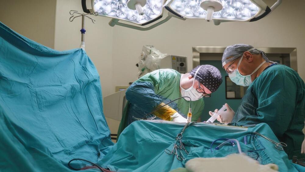
Four astronauts aboard the orbiting lab practiced surgical techniques to heal wounds in microgravity on Friday in the Kibo laboratory module. The quartet split up in groups of two with NASA astronaut Bob Hines joining ESA (European Space Agency) Flight Engineer Samantha Cristoforetti for the first practice session during the morning. In the afternoon, NASA Flight Engineers Kjell Lindgren and Jessica Watkins began their session studying how to take biopsies and suture wounds inside the Life Science Glovebox.

During the middle of the day, the foursome had time set aside time for gathering frozen research samples inside science freezers and preparing them for departure back to Earth inside the SpaceX Dragon resupply ship. Dragon is due to leave the station on Aug. 18 loaded with over 4,000 pounds of station supplies and science experiments after 33 days docked to the Harmony module’s forward port. The commercial cargo craft will parachute to a splashdown off the coast of Florida the next day for retrieval by NASA and SpaceX personnel.

Human Augmentation Examples. What are examples of augmentations to the human body? Should we allow such augmentation methods and technologies? What are dangers, and risks involved? How can society benefit from these developments?

On Brave New World conference 2020 I gave this webinar with the title: ‘The Human Body. The Next Frontier’.

Please leave a comment if you like the video or when you have a question!

Content:
0:00 Start.
0:29 Introduction by moderator Jim Stolze.
1:05 My story.
2:54 Jetson Fallacy (by professor Michael Bess)
3:43 Examples: data, genetic modification, and neuroscience.
6:09 Benefits.
7:09 Risks and dangers.
8:28 Stakeholders: companies, countries, and military organizations.
11:54 Solutions: regulation, debate, and stories.
14:42 My conclusion.
15:03 End.

🙌 Hire me:
Keynote or webinar: https://peterjoosten.org.

🚀 Want to know more:

A group of scientists from the University College London has developed an artificial intelligence (AI) algorithm that can detect drug-resistant focal cortical dysplasia (FCD), a subtle anomaly in the brain that leads to epileptic seizures. This is a promising step for scientists toward detecting and curing epilepsy in its early stages.

To develop the algorithm, the Multicentre Epilepsy Lesion Detection project (MELD) gathered more than 1,000 patients’ MRI scans from 22 international epilepsy centers, which reports where anomalies are in cases of drug-resistant focal cortical dysplasia (FCD), a major reason behind epilepsy.

Specific proteins in prokaryotes detect viruses in unexpectedly direct ways.

Bacteria use a variety of defense strategies to fight off viral infection. STAND ATPases in humans are known to respond to bacterial infections by inducing programmed cell death in infected cells. Scientists predict that many more antiviral weapons will be discovered in the microbial world in the future. Scientists have discovered a new unexplored microbial defense system in bacteria.

Researchers uncovered specific proteins in prokaryotes (bacteria and archaea) that detect viruses in unexpectedly direct ways, recognizing critical parts of the viruses and causing the single-celled organisms to commit suicide to stop the infection within a microbial community, according to a press release published in the official website of the Massachusetts Institute of Technology (MIT) on Thursday.

The discovery was made by a team of scientists led by researchers at the Broad Institute of MIT and Harvard and the McGovern Institute for Brain Research at MIT.

“This work demonstrates a remarkable unity in how pattern recognition occurs across very different organisms,” said Feng Zhang, senior author and James, and Patricia Poitras Professor of Neuroscience at MIT.

“It’s been very exciting to integrate genetics, bioinformatics, biochemistry, and structural biology approaches in one study to understand this fascinating molecular system.”

Bacteria use a variety of defense strategies to fight off viral infection, and some of these systems have led to groundbreaking technologies, such as CRISPR-based gene editing.

Monocytes, a type of white blood cell, are alone capable of facilitating faster wound healing, says study.

Scientists from the University of Calgary, Canada, have discovered a promising new approach to treating bacterial skin infections. The research showed that monocytes alone are capable of facilitating faster wound healing. The researchers’ next step is to better understand how immune cells like neutrophils function during infection. Researchers have discovered a promising new approach to treating bacterial skin infections.

A team of scientists from the University of Calgary, Canada, revealed new insights which could lead to advancements in the treatment of bacterial infections and wounds, according to a study published in Nature science journal on Friday.

“It is exciting that we have made a fundamental discovery that could improve infections and tissue repair in humans, especially hard-to-treat cases,” said the study’s first author Dr. Rachel Kratofi in the press release. “Translating our research from bench to bedside will require many more experiments and involve a model more closely related to human disease.”

The research showed that monocytes alone are capable of facilitating faster wound healing. Monocytes contribute to wound healing by regulating leptin levels and blood vessel growth. They also produce ghrelin, a hormone that aids in wound healing. Historically, researchers believed that neutrophils and monocytes (white blood cells) were both recruited to clear bacteria from an infected site on the skin. When these cells work together, they serve as our bodies first line of defense against the immune system. The connection between metabolic hormones and tissue repair Ghrelin is produced by the stomach when you are hungry, whereas leptin is produced by fat cells after you eat a meal and feel full. This ghrelin-leptin balance has long been recognized as important for metabolism and diet. Still, its relationship to immune mechanisms and tissue repair has been unknown until now. Kratofil was able to visualize the immune response to Staphylococcus aureus (S. aureus) bacteria in an animal model using intravital microscopy, which allows observation of live cells and is a specialization of the university’s Kubes Lab.

Full Story:


This article tells of possible way to increase brain intelligence through a certain mutation which in theory could be altered for biological singularity like effects in the future.


Humans carrying the CORD7 (cone-rod dystrophy 7) mutation possess increased verbal IQ and working memory. This autosomal dominant syndrome is caused by the single-amino acid R844H exchange (human numbering) located in the 310 helix of the C2A domain of RIMS1/RIM1 (Rab3-interacting molecule 1). RIM is an evolutionarily conserved multi-domain protein and essential component of presynaptic active zones, which is centrally involved in fast, Ca2+-triggered neurotransmitter release. How the CORD7 mutation affects synaptic function has remained unclear thus far. Here, we established Drosophila melanogaster as a disease model for clarifying the effects of the CORD7 mutation on RIM function and synaptic vesicle release.

To this end, using protein expression and X-ray crystallography, we solved the molecular structure of the Drosophila C2A domain at 1.92 Å resolution and by comparison to its mammalian homolog ascertained that the location of the CORD7 mutation is structurally conserved in fly RIM. Further, CRISPR/Cas9-assisted genomic engineering was employed for the generation of rim alleles encoding the R915H CORD7 exchange or R915E, R916E substitutions (fly numbering) to effect local charge reversal at the 310 helix. Through electrophysiological characterization by two-electrode voltage clamp and focal recordings we determined that the CORD7 mutation exerts a semi-dominant rather than a dominant effect on synaptic transmission resulting in faster, more efficient synaptic release and increased size of the readily releasable pool but decreased sensitivity for the fast calcium chelator BAPTA.

Circa 2015


“Their work has provided fundamental knowledge of how a living cell functions and is, for instance, used for the development of new cancer treatments,” the Royal Swedish Academy of Sciences said.

Thousands of alterations to a cell’s genome occur every day due to spontaneous changes and damage by radiation, free radicals and carcinogenic substances — yet DNA remains astonishingly intact.

To keep genetic materials from disintegrating, a range of molecular systems monitor and repair DNA, in processes that the three award-winning scientists helped map out.