This website uses a security service to protect against malicious bots. This page is displayed while the website verifies you are not a bot.

The threat is to the librarian. The threat is to the small, vanishing population of people who still go into the hexagons. Who still pull a book from the shelf. Who still spend three days reading it. Who still close it and feel changed. That practice is not a hobby. It is a technology. One older than print, older than the codex, possibly older than writing. It is a process of assembly inside one human skull. The kind of patient, sequential, focused and embodied attention that produces what we used to call understanding. AI does not produce that attention. AI produces a feeling that closely resembles attention while being something else, the way saccharin produces a feeling that closely resembles sweetness while being something else.
If this practice disappears, the Library will not notice. The books will not notice. The infinite hexagons will continue to extend in every direction. There will be no one in them. There will only be the queries, falling into the air, decaying into training data, generating fresh continuations for an audience that no longer reads them. Only, occasionally, glances at a summary.
This is the message Borges was telegraphing. This is what he saw, sitting in the National Library of Argentina, going slowly blind, surrounded by more books than any one man could read. He saw that the deepest threat to a literary culture was not the burning of books. It was the rendering of books unnecessary. He saw that a Library of Babel which contained every possible answer was, paradoxically, the most efficient instrument ever conceived for ending the practice of reading. And he saw, finally, that the only response available to a serious person was the response his narrator chose. To stop searching for the catalogue of catalogues. To return to one’s own hexagon. To pick up one particular book. To read it slowly. To die, eventually, a few leagues from where one was born, with one’s body falling through the fathomless air.
Join us on Patreon! / michaellustgartenphd.
Discount Links/Affiliates:
Blood testing (where I get the majority of my labs, for those who blood test with Quest): https://www.ultalabtests.com/partners… those who blood test with LabCorp: https://www.anrdoezrs.net/click-10161… At-Home Metabolomics: https://www.iollo.com?ref=michael-lus… Use Code: CONQUERAGING At Checkout Clearly Filtered Water Filter: https://get.aspr.app/SHoPY Epigenetic, Telomere Testing: https://trudiagnostic.com/?irclickid=… Use Code: CONQUERAGING NAD+ Quantification: https://www.jinfiniti.com/intracellul… Use Code: ConquerAging At Checkout Oral Microbiome: https://www.bristlehealth.com/?ref=mi… Enter Code: ConquerAging SiphoxHealth Blood Testing (ApoB, GrimAge): https://siphoxhealth.com/mlustgarten Green Tea: https://www.ochaandco.com/?ref=fqbtflod Use Code: ML10OFF Diet Tracking: https://shareasale.com/r.cfm?b=139013… If you’d like to support the channel, you can do that with the website, Buy Me A Coffee: https://www.buymeacoffee.com/mlhnrca Conquer Aging Or Die Trying Merch! https://my-store-d4e7df.creator-sprin… George’s YT channel: / @reprogrampodcast The Murphy Lab website: https://murphylaboratory.com/ X: @DrGJMurphy.
Blood Testing Essentials (Biological Age, CVD-Risk, Kidney Health and Function):
PhenoAge (Biological Age): https://www.ultalabtests.com/partners…
Risk-weighted ApoB (a better CVD predictor than LDL, non-HDL cholesterol, and ApoB): https://www.ultalabtests.com/partners…
Kidney health and function: https://www.ultalabtests.com/partners…
Late last month, Jurassic Park actor Sam Neill put the treatment in the spotlight, revealing his stage three cancer was in remission after undergoing CAR T-cell therapy as part of a clinical trial in Sydney. He stopped short of describing his remission as a miracle – the success, he said, was “science at its best”
The history of CAR (for “chimeric antigen receptor”) T-cell therapy is one of small discoveries accumulating over decades, leading to major advances in patient care. Pioneered in the 1990s, the therapy has exploded in the past decade. Four CAR T-cell therapies have been approved by the Therapeutic Goods Administration for use in Australia since 2018. All are for blood cancers.
The success of those therapies is increasing enthusiasm among researchers and clinicians that CAR T-cell therapies will soon become a major weapon in the battle against cancer. It is now being tweaked to combat solid tumours, with promising early signs of success tempered by the difficulties in tailoring T-cells to find their target. The future may even see it become an injectable.
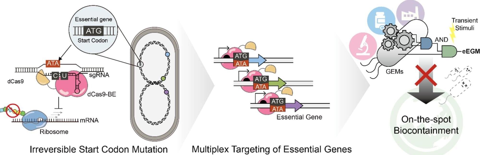
Engineered microorganisms are widely used in industrial biotechnology and biopharmaceutical applications, including the production of biofuels, sustainable chemicals, and therapeutic compounds. However, concerns remain regarding the unintended environmental release and uncontrolled proliferation of genetically engineered microbes. For this reason, biocontainment technologies, which are designed to prevent microorganisms from surviving outside controlled environments, have become increasingly important in both academia and industry.
Conventional biocontainment strategies have relied on auxotrophy-based approaches, toxin–antitoxin systems, or DNA cleavage-based technologies such as CRISPR-Cas9. However, these methods often suffer from environmental dependency, genetic instability, and the risk of unintended mutations and cellular stress caused by DNA double-strand breaks.
In particular, DNA cleavage-based systems may compromise genomic stability and allow certain mutant cells to escape survival control. In addition, CRISPR interference (CRISPRi)-based systems are inherently reversible, posing challenges for achieving complete and permanent control of cell viability.
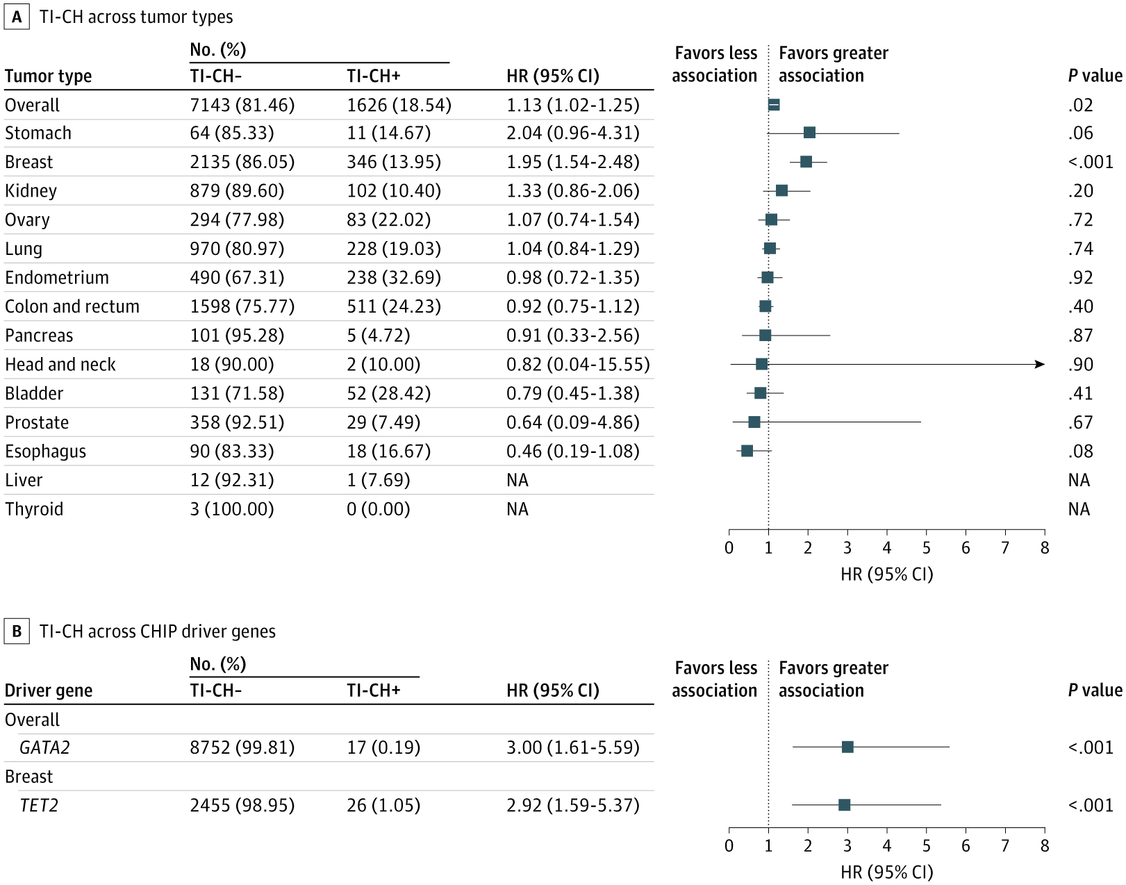
Tumor-infiltrating clonal hematopoiesis was detected in 18% of patients with solid tumors and associated with older age, prior cytotoxic chemotherapy, and reduced overall survival, especially in breast cancer.
This retrospective cohort study investigated the association of TI-CH with clinical factors and its impact on OS in patients with solid tumors. The prevalence of TI-CH in this patient cohort was higher than in treatment-naive cohorts but lower than that in cohorts with higher rates of cytotoxic chemotherapy and radiotherapy. In addition, the prevalence of TI-CH was higher in patients with MSI-high colorectal tumors than in those with MSS colorectal tumors. Analysis of clinical factors revealed that each decade of increasing age and a history of cytotoxic chemotherapy were significantly associated with higher odds of TI-CH. Although TI-CH was associated with worse OS in the whole cohort (pan-cancer analysis), this outcome was most pronounced in patients with breast tumors. Furthermore, TI-CH of GATA2 in the whole cohort and TI-CH of TET2 in patients with breast tumors had the most prominent associations with worse OS.
The accumulation of somatic variants in hematopoietic stem cells with age provides a competitive advantage, leading to CHIP.2 Additionally, cytotoxic chemotherapy induces gene-specific clonal expansion by allowing clones with variants in DNA damage response genes (eg, TP53, PPM1D) to outcompete other clones because such variants are associated with chemoresistance.25 The TI-CH prevalence in our study was intermediate between treatment-naive and treatment-experienced cohorts. It was higher than in the former due to prior therapy and lower than in the latter owing to reduced exposure to cytotoxic chemotherapy and radiotherapy. This finding is notable given this study cohort’s older age, a known factor for increasing CHIP prevalence.6, 7 Furthermore, we found that TI-CH prevalence was higher in patients with MSI-high colorectal tumors than in those with MSS colorectal tumors. To our knowledge, this finding has not been previously reported.
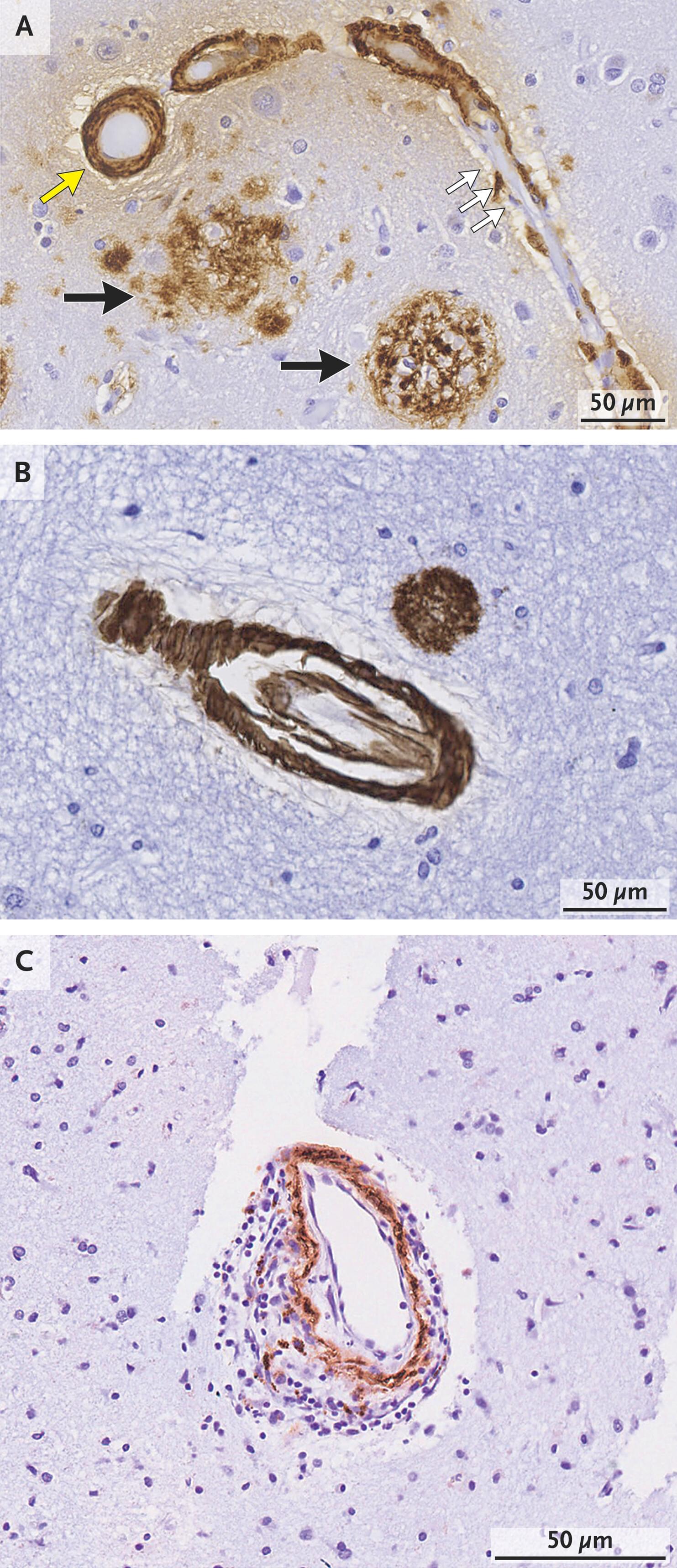
Cerebral amyloid angiopathy is a major cause of hemorrhagic stroke, a frequent contributor to age-related cognitive impairment, and a key component in adverse responses to beta-amyloid (Aβ) immunotherapy. Defined by pathological deposition of Aβ in the small blood vessels of the brain, cerebral amyloid angiopathy is most often diagnosed on the basis of magnetic resonance imaging studies showing multiple hemorrhages or leptomeningeal blood products within or overlying the cerebral cortex. The disorder typically manifests as hemorrhagic stroke or as a contributing factor to cognitive decline and, less commonly, with transient focal neurologic symptoms or a cerebral inflammatory autoimmune syndrome.
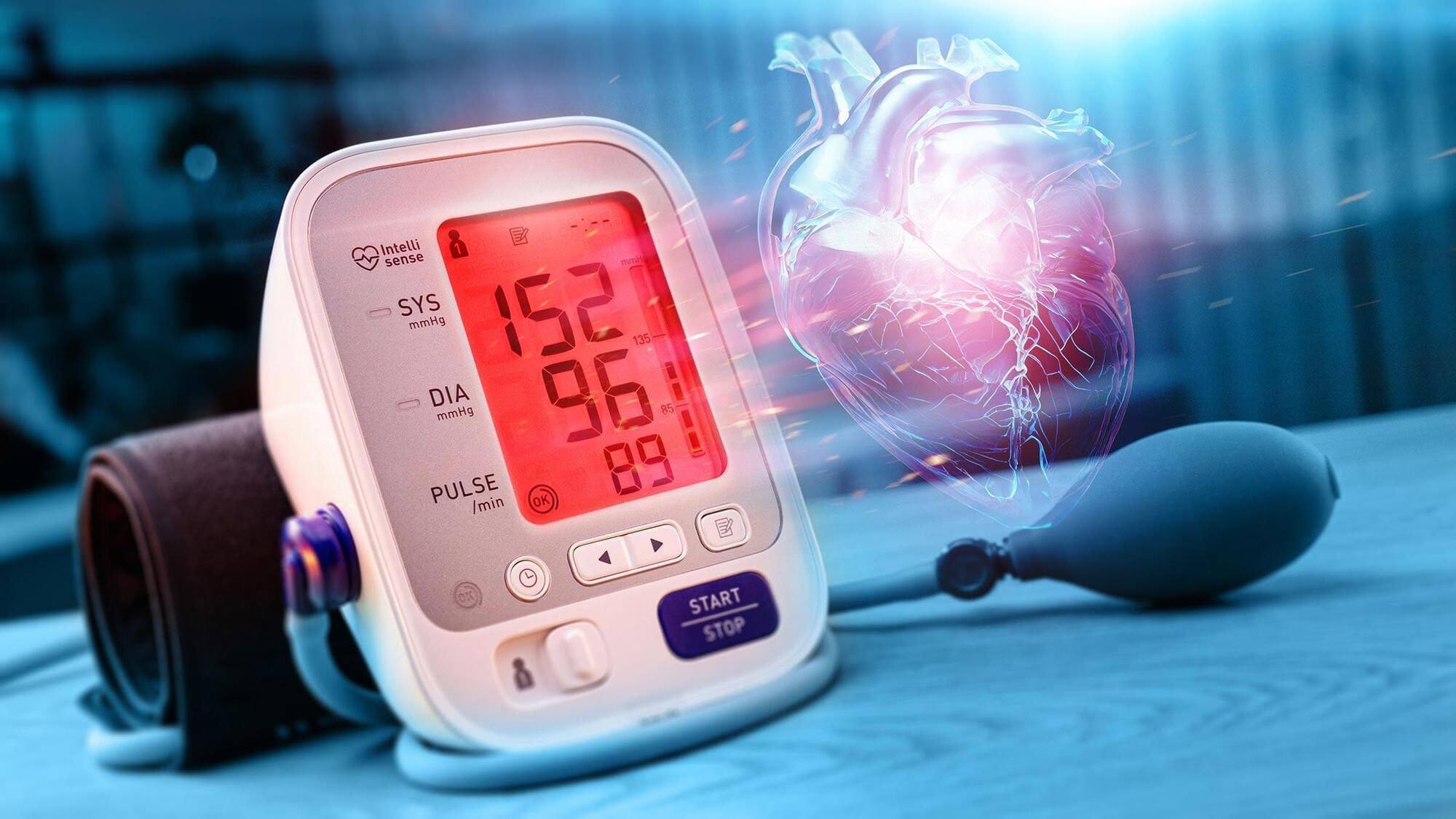
Scientists in Japan say a common supplement may actually help “unclog” certain diseased heart arteries from the inside out.
A simple food supplement sold in Japan may have helped reverse a dangerous form of heart disease that often resists standard treatment, according to researchers at Osaka University. The findings, originally published in the European Heart Journal, continue to attract attention because they describe something rarely seen in cardiology: clogged heart arteries becoming noticeably clearer after a nutritional intervention rather than conventional cholesterol lowering alone.
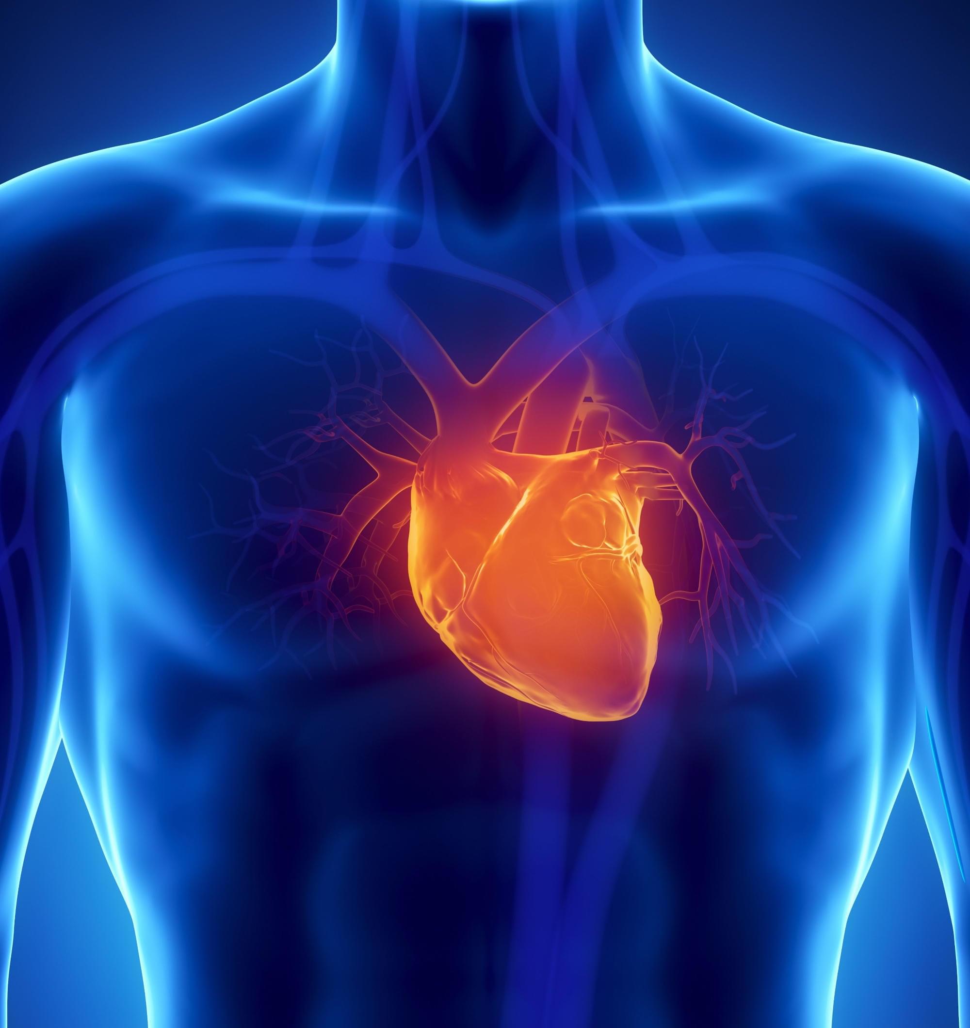
Scientists target a hidden form of heart disease.