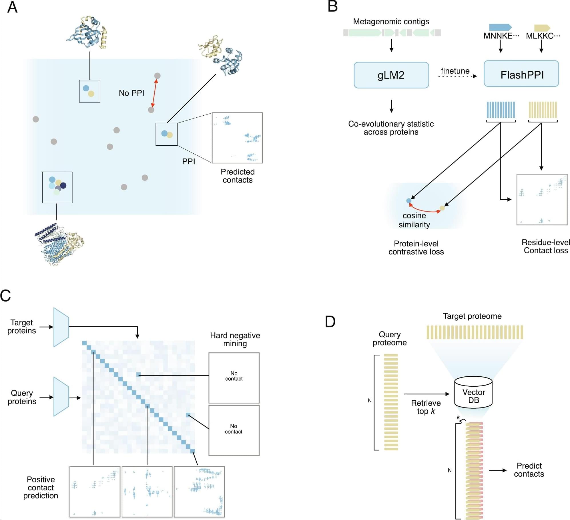
Protein–protein interactions (PPIs) underpin biological function, yet proteome-scale interaction prediction remains bottlenecked by the quadratic computational complexity of all-vs.-all pairwise comparisons. Here, we present FlashPPI, a contrastive learning framework, grounded in residue-level interactions, that enables linear-time prediction of physical protein interfaces across a microbial proteome. By leveraging a genomic language model that captures cross-protein coevolutionary signals from metagenomic sequences, FlashPPI aligns interacting partners in a shared latent space. We demonstrate a four-fold performance increase over existing sequence-based methods, while reducing proteome-wide screening time from days to minutes. Crucially, FlashPPI achieves comparable screening performance to state-of-the-art structure-folding models at a fraction of the computational cost. Finally, we integrate FlashPPI into an interactive web platform that combines predicted networks with functional annotations and genomic context, making proteome-wide network analysis rapid and accessible for microbial discovery.
Not to toot my own horn or anything, but I can extend my empathy beyond myself just enough to imagine someone else’s perspective, fully knowing I’ll never completely understand the texture of their experience. But as a right-handed person, I will never, ever be able to do that for left-handed people. There’s just something in my brain preventing me from understanding how someone can navigate the world primarily using the hand I mostly rely on to accidentally test the sharpness of kitchen knives.
So naturally, it always made sense to me that around 90 percent of humans are right-handed. What never made sense was why. According to new research published in PLOS Biology, we may have finally figured it out: humans became overwhelmingly right-handed because we started walking upright and developed massive brains.
Researchers from the University of Oxford analyzed more than 2,000 primates across 41 species, comparing handedness with factors like social behavior, diet, body size, and movement. Nothing fully explained humanity’s innate steadfast dedication to right-handedness until researchers started factoring in brain size and the ratio between leg and arm length.
A research team has developed a methodology to precisely design and control the “degree of disorder” in nanopattern arrays using metal-infiltrated block copolymer (BCP) thin films. The work was led by Professor So Youn Kim of the Seoul National University College of Engineering Department of Chemical and Biological Engineering, in collaboration with Professor Su-Mi Hur’s team at DGIST and Professor S. Joon Kwon’s team at Sungkyunkwan University. The paper is published in the journal Nature Communications. The study was selected as an Editors’ Highlight in materials science and chemistry.
This disordered nanopattern fabrication technology is regarded as an innovative approach that enables precise control of nanoscale disorder structures—previously difficult to regulate—thereby opening new possibilities in the design of nano-optical and nanoelectronic devices.
In ordered structures, waves propagate over long distances, whereas in disordered structures, repeated scattering can lead to localization, where waves remain confined within a specific region. Such disordered structures exhibit unique functionalities that can induce localization phenomena for various types of waves, including light, sound and heat.
An array of artificial neurons printed on a flexible wafer produce spiking waveforms closely matching biological action — and drive living mouse brain cells to fire.
From cosmic rays to solar storms, space travel is a radiation gauntlet… but water may be the simplest, smartest solution. Discover how future starships might turn their life-support systems into life-saving armor.
Get Nebula using my link for 50% off an annual subscription: https://go.nebula.tv/isaacarthur. Watch my exclusive video Nearby Supernovae: https://nebula.tv/videos/isaacarthur–… out Gods & Monsters: https://nebula.tv/curiousarchive/gods… 🛒 SFIA Merchandise: https://isaac-arthur-shop.fourthwall… 🌐 Visit our Website: http://www.isaacarthur.net ❤️ Support us on Patreon: / isaacarthur ⭐ Support us on Subscribestar: https://www.subscribestar.com/isaac-a… 👥 Facebook Group: / 1,583,992,725,237,264 📣 Reddit Community: / isaacarthur 🐦 Follow on Twitter / X: / isaac_a_arthur 💬 SFIA Discord Server: / discord Credits: Fishbowl Starships Water As Shielding Episode 721; June 1, 2025; Nebula Exclusive Written, Produced, & Narrated by: Isaac Arthur Graphics: Bryan Versteeg, Jeremy Jozwik, Udo Schroeter Select imagery/video supplied by Getty Images Music Courtesy of Epidemic Sound http://epidemicsound.com/creator Taras Harkavyi, “Alpha and…” Chris Zabriskie, “Unfoldment, Revealment”, “A New Day in a New Sector” “Oxygen Garden”, “Wonder Cycle” Stellardrone, “Red Giant”, “Billions and Billions” Chapters: 0:00 Intro 1:45 The Threat — Radiation In Space 2:36 Galactic Cosmic Rays (GCRs) 3:44 Solar Particle Events (SPEs) 4:16 Van Allen Belt Radiation 5:19 Radiation’s Impact on Humans and Equipment 8:18 Radiation Shielding Basics 9:18 Water as a Radiation Shield 11:19 Effectiveness of Water 15:42 Difficulties Using Water 17:29 Beyond Water: Alternative Radiation Shielding Methods 17:59 Metallic Shielding 18:58 Regolith & Asteroid-Based Shielding 20:27 Hydrogen-Rich Polymers 21:22 Graphene, CNTs, and BNNTs 23:54 Active Shielding: Magnetic & Plasma Barriers 24:49 Fusion Fuel Shielding 25:27 Hybrid Shielding Approaches 28:16 God & Monsters 29:26 The Future of Radiation Shielding 30:00 Smart & Self-Healing Shielding 31:29 Artificial Magnetospheres 33:22 Biological Adaptation 35:35 Radiation-Resistant AI & Robotics 37:45 The Future of Space Radiation Protection 38:55 The Future of Water-Based Shielding. Check out Gods \& Monsters: https://nebula.tv/curiousarchive/gods…
🛒 SFIA Merchandise: https://isaac-arthur-shop.fourthwall… 🌐 Visit our Website: http://www.isaacarthur.net. ❤️ Support us on Patreon: / isaacarthur. ⭐ Support us on Subscribestar: https://www.subscribestar.com/isaac-a… 👥 Facebook Group: / 1583992725237264 📣 Reddit Community: / isaacarthur. 🐦 Follow on Twitter / X: / isaac_a_arthur. 💬 SFIA Discord Server: / discord. Credits: Fishbowl Starships Water As Shielding. Episode 721; June 1, 2025; Nebula Exclusive. Written, Produced, \& Narrated by: Isaac Arthur. Graphics: Bryan Versteeg, Jeremy Jozwik, Udo Schroeter. Select imagery/video supplied by Getty Images. Music Courtesy of Epidemic Sound http://epidemicsound.com/creator. Taras Harkavyi, \
Water is the most studied molecule on Earth, yet a surprisingly basic question has gone unanswered for decades: When water is squeezed into gaps just a few molecules wide—as happens inside nanoscale pores, membranes and biological channels—does it become more or less chemically reactive?
This matters because water’s most fundamental chemical property is its ability to split into two charged species, H₃O⁺ (the hydronium ion) and OH⁻ (the hydroxide ion). This reaction defines the pH, a measure of how acidic or alkaline (basic) a solution is, and underpins all of acid-base chemistry, from how enzymes work in your cells to how electrodes function in batteries.
Through this research, the scientists wanted to understand whether (and how) confining water to nanometer-scale spaces affects this behavior.
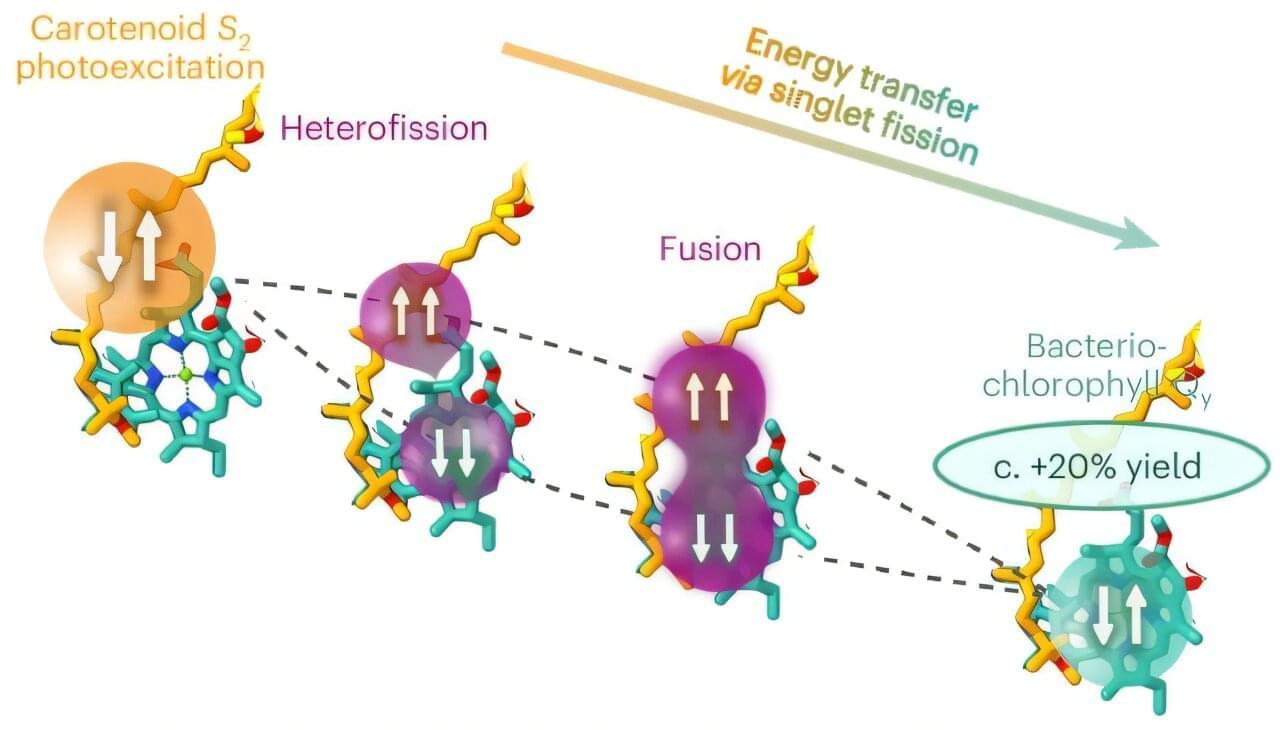
Researchers have discovered how certain photosynthetic bacteria use a sophisticated quantum mechanism to increase their efficiency when capturing sunlight. The study, published today in the journal Nature Chemistry and led by Professor Jenny Clark, reveals that nature has been using a process called “singlet fission,” effectively a “two-for-one” energy deal, to optimize solar harvesting. The findings provide a new blueprint for green technology, particularly as engineers attempt to copy this mechanism to build next-generation solar panels and quantum technologies.
While scientists have long understood the basic rules of how plants and bacteria convert light into chemical fuel, the biological role of singlet fission has historically remained poorly understood.
Over the past decade, Professor L. Mahadevan’s Soft Math Lab at the Harvard John A. Paulson School of Engineering and Applied Sciences (SEAS) has helped establish how the ancient Japanese paper arts of folding or cutting can be used to inversely design structures that transform dramatically in shape and function. Now, the researchers have created a new class of shape-changing matter, based not on folds or cuts, but linkages—networks of interconnected scissor mechanisms that collapse into lines and deploy into curved surfaces.
The study published in the Proceedings of the National Academy of Sciences, led by physics graduate student Noah Toyonaga, establishes a mathematical and physical framework for what the authors call collapsible scissored surfaces—deployable lattices of two-bar linkages that can transform from a one-dimensional collapsed state into two-dimensional structures with prescribed geometry.
“Origami showed how folds can encode shape,” said senior author Mahadevan, the Lola England de Valpine Professor of Applied Mathematics, of Organismic and Evolutionary Biology, and of Physics. “Kirigami showed how cuts can unlock motion and functionality. This work asks a complementary question: What can be achieved when the basic building block is not a fold or a cut, but a linkage?”
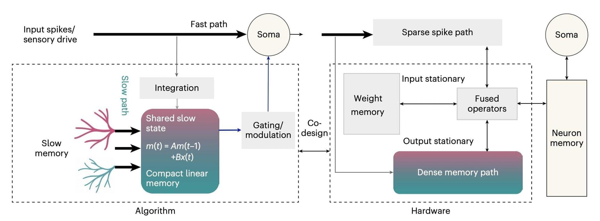
Spiking neural networks (SNNs) are artificial intelligence (AI) models inspired by how biological neurons communicate with each other. While biological neurons exchange information in the form of electrical impulses, SNNs rely on brief signals known as spikes.
SNNs have proved promising for reducing power consumption, as developers can ensure they do not process information continuously, but rather only when meaningful changes occur. This could be highly advantageous, as current AI systems are known to consume large amounts of energy.
While some SNNs introduced in the past achieved encouraging results, they typically struggle to retain useful information (i.e., context) for long periods. This was found to be particularly challenging when the models have only a limited amount of data storage available or are operating under energy constraints.
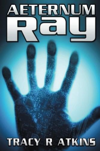
That was Tracy R. Atkins’ message when I sat down with him 14 years ago, and it lands harder now than it did then.
While almost every story about #ArtificialIntelligence was busy imagining the apocalypse, Tracy wrote a novel that flatly refused to. Aeternum Ray is unapologetically utopian: a series of letters from a 240-year-old father to his newborn son, looking back across centuries of love, loss, and a world watched over by an AI named Ray.
In our conversation, we get into what the #Singularity actually means to him, why he chose to write utopia when dystopia sells, whether humanity’s future is digital or whether biology still matters, and the uncomfortable question of whether we even survive the road to get there.
Fourteen years on, the technology has caught up to much of what we talked about. The harder question is whether our reasons for building it have, and that is the part I keep coming back to.
So is openly imagining a good future naive, or is it the most radical thing a #futurist can do? Watch the interview and decide for yourself.