👉For business inquiries: [email protected].
✅ Instagram: https://www.instagram.com/pro_robots.
In the quest to overcome the limitations of the human body and mind, scientists worldwide are diligently working on various technologies. The question arises: What will human beings become after undergoing numerous enhancements? Will we retain our identity while embracing the possibilities offered by artificial intelligence? What extraordinary capabilities will biotechnology bestow upon us? And how will our emotions and desires evolve as our bodies undergo transformation?
Join us on a captivating journey to the year 2050, as we delve into the frontiers of scientific research, consult with visionary futurists, and examine the predictions of brilliant minds. Together, we will explore the profound changes that lie ahead!
00.00 — Introduction.
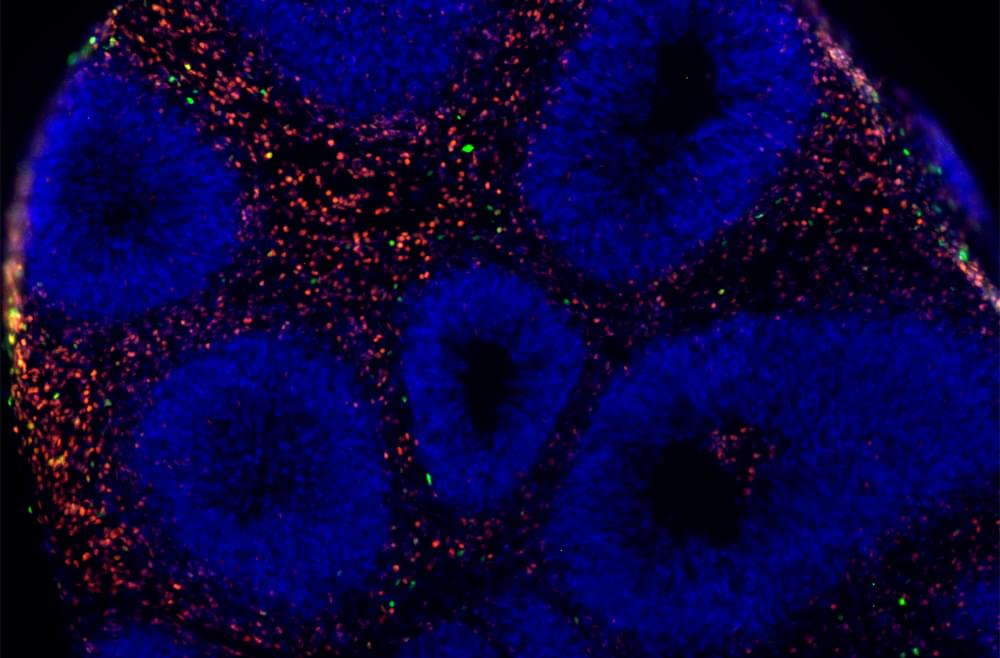
01:15 — Matrix-Like Innovation: Baby-Growing Factories Bring Science Fiction to Reality.
02:33 — The Future of Longevity: Exploring Eternal Youth Technologies.
03:51 — Unlocking Superpowers: Genetic Engineering Takes Humans and Animals to New Heights!
05:11 — Brain Implants in 2050: The Future of Communication, Control, and Enhanced Human Abilities.
Redefining Human Life.
In the year 2050, the human body will undergo a transformation like never before. For the first time in our 300,000-year history, evolution will not solely rely on natural selection but rather on deliberate re-engineering through technology.
Revolutionary Childbirth.