“This research sheds light on an important facet of modern Mars: the interaction of the atmosphere and the surface,” said Dr. Paul Byrne. [ https://www.labroots.com/trending/space/30400/martian-dust-s…eactions-2](https://www.labroots.com/trending/space/30400/martian-dust-s…eactions-2)
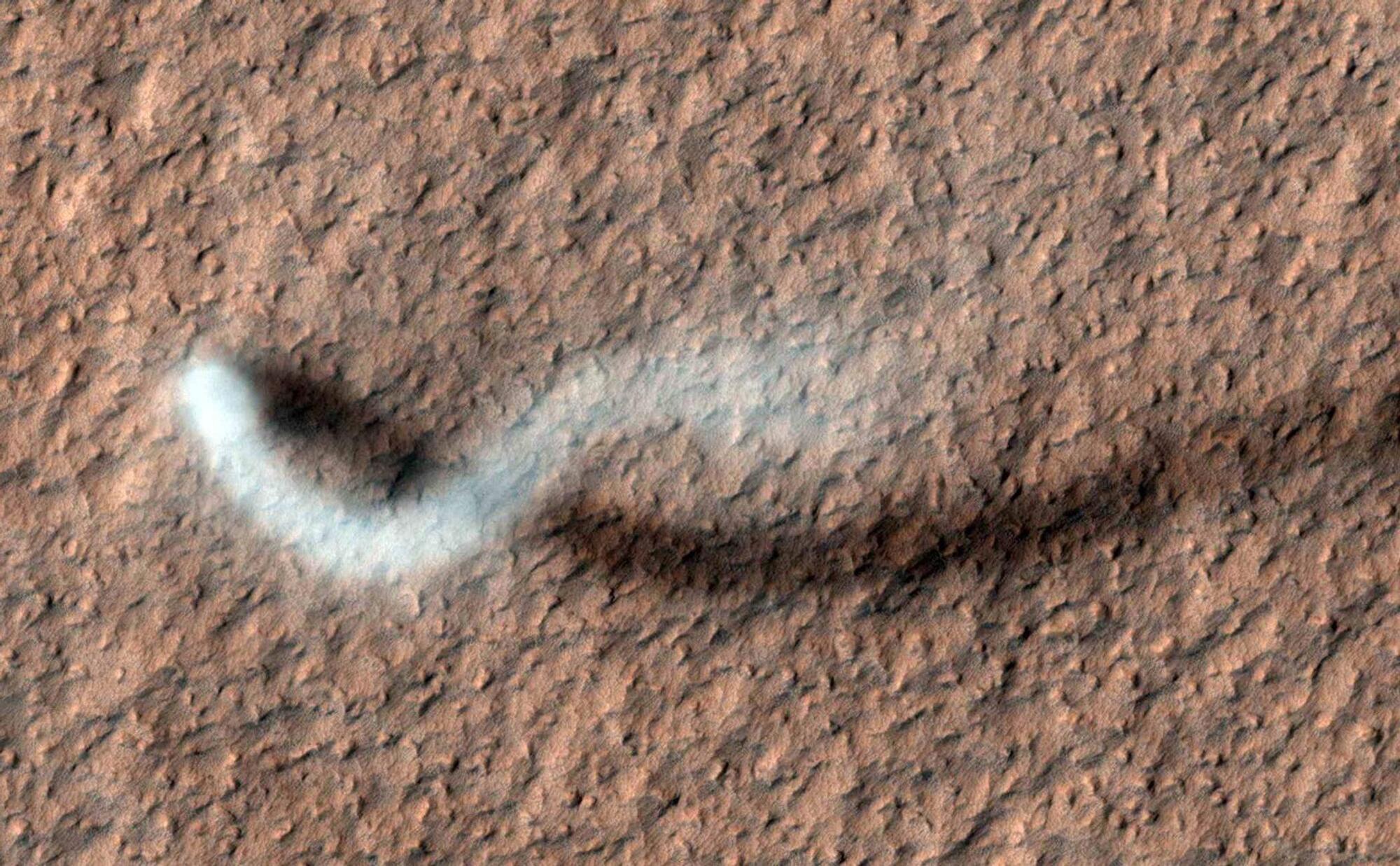
How does static electricity shape the surface of Mars? This is what a recent study published in Earth and Planetary Science Letters hopes to address as an international team of scientists investigated atmosphere-surface interactions on Mars, specifically regarding electrostatic discharge, or static electricity. This study has the potential to help scientists better understand atmosphere-surface interactions on planetary bodies and how this could help find life beyond Earth.
For the study, the researchers conducted a series of laboratory experiments to simulate how dust storms and dust devils on Mars could trigger the production of compounds like perchlorates and carbonates within the Martian regolith (often mistakenly called “soil”) and hydrochloric acid (HCl) in the atmosphere. The motivation for the study was to gain insight into how planets work, specifically regarding their geological activity.
In the end, the researchers found that static electricity from Martian dust activities are responsible for producing perchlorates and carbonates in the Martian regolith and HCl in the Martian atmosphere. The study’s results were compared with real-world data obtained from the European Space Agency’s ExoMars Trace Gas Orbiter and NASA’s Curiosity rover for atmospheric and surface data, respectively.