A team in China figured out a way to take control of a rat and “steer” it through a maze with their thoughts.


Virgin Galactic sent three human beings on Unity for the first time in Friday’s supersonic test flight, which reached three times the speed of sound on its way up. Just before the flight, Richard Branson’s space tourism company told CNBC that astronaut trainer Beth Moses is on the company’s spacecraft Unity, along with the two pilots.
“Beth Moses is on board as a crew member,” a Virgin Galactic spokeswoman told CNBC. “She will be doing validation of some of the cabin design elements.”

I wonder how diet mixed with CBD oil treatment can work on neurodegenerative diseases? A man has told of how he “got his mum back” after a diagnosis of Alzheimer’s disease, in part, by getting her to follow a diet high in berries and leafy green vegetables.
One man has ‘got his mum back’ from the ravages of Alzheimer’s, partly…

#ClinicalTrial The most common syndrome in patients with severe dementia is agitated behavior, which is often characterized by a combination of violent behavior (physical or verbal), restlessness, and inappropriate loudness. The treatment options for this syndrome are limited and lead to severe side effects. In vivo experiments on animals and clinical studies on adults show that cannabinoids could have a beneficial effect on behavioral disorders in general, and in dementia-related disorders in particular.
Full Text View.

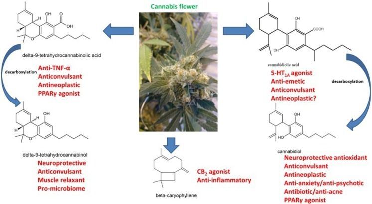
Neurological therapeutics have been hampered by its inability to advance beyond symptomatic treatment of neurodegenerative disorders into the realm of actual palliation, arrest or reversal of the attendant pathological processes. While cannabis-based medicines have demonstrated safety, efficacy and consistency sufficient for regulatory approval in spasticity in multiple sclerosis (MS), and in Dravet and Lennox-Gastaut Syndromes (LGS), many therapeutic challenges remain. This review will examine the intriguing promise that recent discoveries regarding cannabis-based medicines offer to neurological therapeutics by incorporating the neutral phytocannabinoids tetrahydrocannabinol (THC), cannabidiol (CBD), their acidic precursors, tetrahydrocannabinolic acid (THCA) and cannabidiolic acid (CBDA), and cannabis terpenoids in the putative treatment of five syndromes, currently labeled recalcitrant to therapeutic success, and wherein improved pharmacological intervention is required: intractable epilepsy, brain tumors, Parkinson disease (PD), Alzheimer disease (AD) and traumatic brain injury (TBI)/chronic traumatic encephalopathy (CTE). Current basic science and clinical investigations support the safety and efficacy of such interventions in treatment of these currently intractable conditions, that in some cases share pathological processes, and the plausibility of interventions that harness endocannabinoid mechanisms, whether mediated via direct activity on CB1 and CB2 (tetrahydrocannabinol, THC, caryophyllene), peroxisome proliferator-activated receptor-gamma (PPARγ; THCA), 5-HT1A (CBD, CBDA) or even nutritional approaches utilizing prebiotics and probiotics. The inherent polypharmaceutical properties of cannabis botanicals offer distinct advantages over the current single-target pharmaceutical model and portend to revolutionize neurological treatment into a new reality of effective interventional and even preventative treatment.
Keywords: cannabis, pain, brain tumor, epilepsy, Alzheimer disease, Parkinson disease, traumatic brain injury, microbiome.
Cannabis burst across the Western medicine horizon after its introduction by William O’Shaughnessy in 1838 (O’Shaughnessy, 1838–1840; Russo, 2017b), who described remarkable successes in treating epilepsy, rheumatic pains, and even universally fatal tetanus with the “new” drug. Cannabis, or “Indian hemp,” was rapidly adopted by European physicians noting benefits on migraine by Clendinning in England (Clendinning, 1843; Russo, 2001) and neuropathic pain, including trigeminal neuralgia by Donovan in Ireland (Donovan, 1845; Russo, 2017b). These developments did not escape notice of the giants of neurology on both sides of the Atlantic, who similarly adopted its use in these indications: Silas Weir Mitchell, Seguin, Gowers and Osler (Mitchell, 1874; Seguin, 1877; Gowers, 1888; Osler and McCrae, 1915).
Imperial researchers have developed a new bioinspired material that interacts with surrounding tissues to promote healing.
Materials are widely used to help heal wounds: Collagen sponges help treat burns and pressure sores, and scaffold-like implants are used to repair broken bones. However, the process of tissue repair changes over time, so scientists are looking to biomaterials that interact with tissues as healing takes place.
Creatures from sea sponges to humans use cell movement to activate healing. Our approach mimics this by using the different cell varieties in wounds to drive healing. Dr Ben Almquist Department of Bioengineering


In my continuing work with the government of UAE / #Dubai, I have an article on #transhumanism that came out in a new portal launched with their recent World Government Summit 2019. Give it a read!
Everywhere around us a “super human” future is rapidly appearing. Sometimes called transhumanism, scientists, programmers, and engineers everywhere are working on radical technologies that not only become a part of our everyday reality, but also fit directly into our bodies.
Some examples are contact lenses that see in the dark. Others are endoskeletons attached to artificial limbs that can lift a half ton of weight. Still others are brain chip implants that read your thoughts and instantly communicate them with others. Sound like science fiction? Indeed, it does. Nevertheless, it’s coming very soon. In fact, much of the technology already exists. Some of it’s being sold commercially at your local superstore or being tested in laboratories right now around the world.

This is Part 3 of a 5-part series by Chogwu Abdul, founder of the Transhumanist Enlightenment Café (TEC), where he explores the thought-provoking intricacies of James Hughes’ “Problems of Transhumanism.”
In this Part, he explores “Liberal Democracy Versus Technocratic Absolutism.”