Toggle light / dark theme

Way cool.


Feb. 2 (UPI) — Scientists at the University of California, Santa Barbara want to study the effects of various mechanical forces on individual brain cells. Until now, however, researchers didn’t have the right tools.

To study brain impacts at the nanoscale, researchers built the world’s tiniest hammer — the μHammer, or “microHammer.” The μHammer is a cellular-scale machine capable of applying a variety of mechanical forces to neural progenitor cells, brain-centric stem cells. Eventually, scientists hope to use the hammer to apply forces to neurons and neural tissue.

The hammer piggybacks on existing cell-sorting technology which isolates individual cells for diagnostics and immunotherapy. Once isolated, the machine can apply a range of forces. Post-impact structural and biomechanical analysis will allow scientists study the effects of focus in near real-time.

A research team led by Professor Ed X. Wu of the Department of Electrical and Electronic Engineering at the University of Hong Kong has used an innovative neuroimaging tool to interrogate the complex brain networks and functions.

The team has successfully manipulated two pioneering technologies: optogenetics and imaging (fMRI), for investigation of the dynamics underlying activity propagation. Their breakthrough to simultaneously capture large-scale brain-wide neural activity propagation and interaction dynamics, while examining their functional roles has taken scientists a step further in unravelling the mysteries of the brain. It could lead to the development of new neurotechnologies for early diagnosis and intervention of brain diseases including autism, Alzheimer’s disease or dementia.

The findings have recently been published in the prestigious international academic journal Proceedings of the National Academy of Sciences (PNAS).

Read more

Interesting read especially as we look at various areas including synbio and super humans.


The results are significant for gene therapy procedures and for our understanding of cell transformation. A team of researchers from the biology department at TU Darmstadt has discovered that the processes for repairing DNA damage are far more complex than previously assumed. The ends of breaks in the double helix are not just joined, they are first changed in a meticulously choreographed process so that the original genetic information can be restored. The results have now been published in the research journal Molecular Cell.

DNA, the carrier of our genetic information, is exposed to continual damage. In the most serious damage of all, the DNA double-strand break, both strands of the double helix are broken and the helix is divided in two. If breaks like this are not efficiently repaired by the cell, important genetic information is lost. This is often accompanied by the death of the cell, or leads to permanent genetic changes and cell transformation. Over the course of evolution, ways to repair this DNA damage have developed, in which many enzymes work together to restore the genetic information with the maximum possible precision.

As it stands today, there are two main ways of repairing DNA double-strand breaks, which differ greatly in terms of their precision and complexity. The apparently simpler method, so-called non-homologous end joining, joins together the break ends as quickly as possible, without placing particular importance on accurately restoring the damaged genetic information. The second method of repair, homologous recombination, on the other hand, uses the exactly identical information present on a sister copy to repair the damaged DNA with great precision. However, such sister copies are only present in dividing cells, as the genetic information has to be duplicated before the cells divide. But most cells in the human body do not undergo division, which therefore assigns them to the apparently more inaccurate method of end joining.

Read more

To generate swarms of new viral particles, a virus hijacks a cell into producing masses of self-assembling cages that are then loaded with the genetic blueprint for the next infection. But the picture of how that DNA is loaded into those viral cages, or capsids, was blurry, especially for two of the most common types of DNA virus on earth, bacterial viruses and human herpesvirus. Jefferson researchers pieced together the three-dimensional atomic structure of a doughnut-shaped protein that acts like a door or ‘portal’ for the DNA to get in and out of the capsid, and have now discovered that this protein begins to transform its structure when it comes into contact with DNA. Their work published in Nature Communications.

“Researchers thought that the portal protein acts as an inert passageway for DNA,” says senior author Gino Cingolani, Ph.D., a Professor in the Department of Biochemistry and Molecular Biology at Thomas Jefferson University and researcher at the Sidney Kimmel Cancer Center. “We have shown that the portal is much more like a sensor that essentially helps measure out an appropriate length of DNA for each capsid particle, ensuring faithful production of new viral particles.”

The finding solves a longstanding puzzle in the field, and reveals a potential drug target for one of the most common human viral pathogens, herpesviruses, which is responsible for diseases such as chicken pox, mononucleosis, lymphomas and Kaposi sarcoma.

Read more

An international team of researchers has identified 83 new DNA changes that strongly determine human height as well as also help predict a person’s risk of developing certain growth disorders.

Height is mostly determined by the information encoded in the human DNA — children from tall parents tend to be taller and those from short parents are shorter.

“Of these 83 genetic variations, some influence adult height by more than 2 cm, which is enormous,” said Guillaume Lettre, Professor at Montreal Heart Institute in Canada.

Read more

Bring to life those old biology and museum specimens back to life. Sort of.

Let’s see Ray Kurzweils prediction of bringing people back from the dead may not be that too far off with this recent discovery. BTW — he may be interested in this one.


(Natural News) Rare animals have been sitting in glass jars on museum shelves across the world for decades, but very little is often known about these specimens. And many people would say that is exactly where they belong: on a shelf, as an object of the past simply to be remembered and admired from afar.

However, a recent scientific breakthrough may be able to breathe new life into these long-extinct species. A new technique for extracting DNA has been developed, and it’s something researchers believe could be used to help bring these long-gone animals into the realm of genomic study. (RELATED: Follow more news about scientific breakthroughs at Scientific.news)

Rare animal specimens are kept in liquid-filled jars as a form of preservation. The liquid preservative, such as formalin, often come with consequences, however. As these creatures are left in the liquid, there is a large amount of DNA fragmenting going on — which makes them less than ideal subjects for DNA extraction.

Read more

Nearly every other year the transistors that power silicon computer chip shrink in size by half and double in performance, enabling our devices to become more mobile and accessible. But what happens when these components can’t get any smaller? George Tulevski researches the unseen and untapped world of nanomaterials. His current work: developing chemical processes to compel billions of carbon nanotubes to assemble themselves into the patterns needed to build circuits, much the same way natural organisms build intricate, diverse and elegant structures. Could they hold the secret to the next generation of computing?

TEDTalks is a daily video podcast of the best talks and performances from the TED Conference, where the world’s leading thinkers and doers give the talk of their lives in 18 minutes (or less). Look for talks on Technology, Entertainment and Design — plus science, business, global issues, the arts and much more.
Find closed captions and translated subtitles in many languages at http://www.ted.com/translate

Follow TED news on Twitter: http://www.twitter.com/tednews
Like TED on Facebook: https://www.facebook.com/TED

Read more

Latest update on the NPL Research on how to have cleaner Quantum Devices.


A paper, based on NPL collaborative research, has been published in the journal Physical Review Letters The work paves the way for the identification and elimination of small amounts of surface defects whose presence on the surfaces of solid state quantum devices is detrimental to their performance.

The research was the result of a fruitful collaboration between NPL’s Quantum Detection Group, the Quantum Device Physics Laboratory at Chalmers University of Technology and the Institute of Chemical Physics at the University of Latvia.

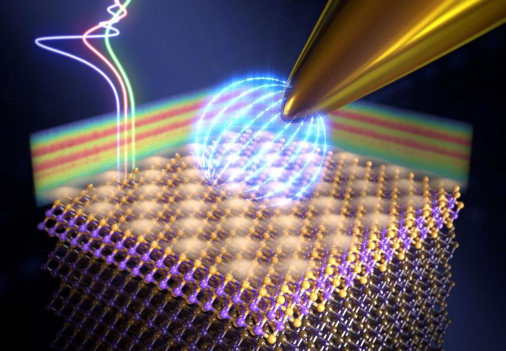
Artistic impression of noise in quantum circuits

The advancement of quantum computing faces a tremendous challenge in improving the reproducibility and robustness of quantum circuits. One of the biggest problems in this field is the presence of noise intrinsic to all these devices, the origin of which has puzzled scientists for many decades.

Read more

I had to take a second review of this since I posted it, and right away I see something quite interesting that folks have overlooked for a while. Will keep you posted.


Scientists funded by the National Institutes of Health have built a new tool to monitor the way cells attach to an adjoining substrate under a microscope.

Analyzing adhesion events can help researchers to understand the way diseases spread, tissues grow, and stem cells differentiate into many specific cell types. The technique provides high-resolution images that can monitor the interactions of cells across longer time periods than previously possible.

Researchers from the University of Illinois at Urbana-Champaign published details of their study in the November 2016 issue of the Progress in Quantum Electronics journal. The researchers demonstrated the method known as photonic crystal-enhanced microscopy (PCEM), on various types of cancer cells and stem cells, revealing that each cell type had its own exclusive adhesion properties.

When it comes to obtaining new energy, solar energy now costs less than fossil fuels, according to a report by the World Economic Forum (WEF). Data from Bloomberg New Energy Finance (BNEF) also show decreased prices, with the mean price of solar power in about 60 countries dropping to $1.65 million per megawatt, closely followed by wind at $1.66 million per megawatt.

Michael Drexler, Head of Long Term Investing, Infrastructure and Development at the World Economic Forum, found the downturn in prices to be an encouraging sign.

“Renewable energy has reached a tipping point—it now constitutes the best chance to reverse global warming. Solar and wind have just become very competitive, and costs continue to fall. It is not only a commercially viable option, but an outright compelling investment opportunity with long-term, stable, inflation-protected returns.”

Read more