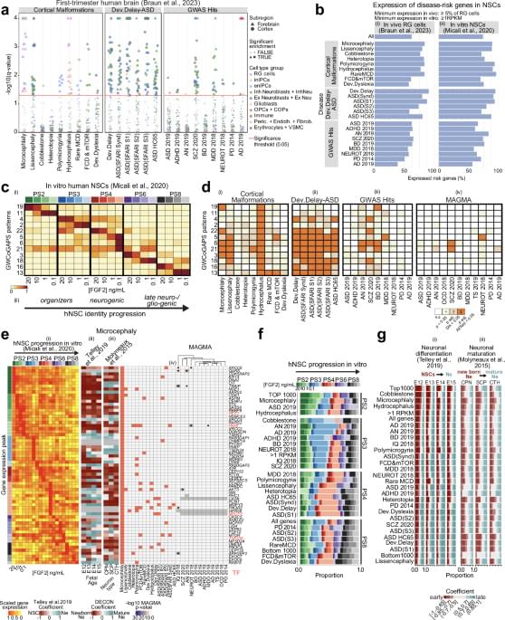
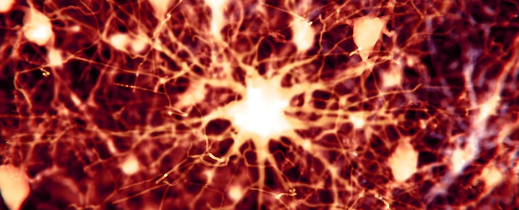
A groundbreaking study has used patient-derived stem cells to recreate a rare genetic form of autism in the lab.
Category: neuroscience – Page 92
A new way of mapping activity and connections between different regions of the brain has revealed fresh insights into how higher order functions like language, thought and attention, are organized.
Traditional models of human brain activity represent interactions in pairs between two different brain regions. This is because modeling methods have not developed sufficiently to describe more complex interactions between multiple regions.
A new approach, developed by researchers at the University of Birmingham is capable of taking signals measured through neuroimaging, and creating accurate models from these to show how different brain regions are contributing to specific functions and behaviors. The results are published in Nature Communications.
Cleveland Clinic Genome Center researchers have unraveled how immune cells called microglia can transform and drive harmful processes like neuroinflammation in Alzheimer’s disease. The study, published in the journal Alzheimer’s & Dementia, also integrates drug databases with real-world patient data to identify FDA-approved drugs that may be repurposed to target disease-associated microglia in Alzheimer’s disease without affecting the healthy type.
The researchers, led by study corresponding author Feixiong Cheng, Ph.D., hope their unique approach of integrating genetic, chemical and human health data to identify drug targets and corresponding drugs will inspire other scientists to take similar approaches in their own research.
Microglia are specialized immune cells that patrol our brains, seeking and responding to tissue damage and external threats like bacteria and viruses. Different types of microglial cells use different methods to keep the brain safe. Some may cause neuroinflammation—inflammation in the brain—to fight invaders or kickstart the repair process in damaged cells. Others may work to “eat” dangerous substances in the brain, and clean up damage and debris. However, during Alzheimer’s disease, new types of microglia can form that promote disease progression.
Chaperone-mediated autophagy (CMA) is the lysosomal degradation of individually selected proteins, independent of vesicle fusion. CMA is a central part of the proteostasis network in vertebrate cells. However, CMA is also a negative regulator of anabolism, and it degrades enzymes required for glycolysis, de novo lipogenesis, and translation at the cytoplasmic ribosome. Recently, CMA has gained attention as a possible modulator of rodent aging. Two mechanistic models have been proposed to explain the relationship between CMA and aging in mice. Both of these models are backed by experimental data, and they are not mutually exclusionary. Model 1, the “Longevity Model,” states that lifespan-extending interventions that decrease signaling through the INS/IGF1 signaling axis also increase CMA, which degrades (and thereby reduces the abundance of) several proteins that negatively regulate vertebrate lifespan, such as MYC, NLRP3, ACLY, and ACSS2. Therefore, enhanced CMA, in early and midlife, is hypothesized to slow the aging process. Model 2, the “Aging Model,” states that changes in lysosomal membrane dynamics with age lead to age-related losses in the essential CMA component LAMP2A, which in turn reduces CMA, contributes to age-related proteostasis collapse, and leads to overaccumulation of proteins that contribute to age-related diseases, such as Alzheimer’s disease, Parkinson’s disease, cancer, atherosclerosis, and sterile inflammation. The objective of this review paper is to comprehensively describe the data in support of both of these explanatory models, and to discuss the strengths and limitations of each.
Chaperone-mediated autophagy (CMA) is a highly selective form of lysosomal proteolysis, where proteins bearing consensus motifs are individually selected for lysosomal degradation (Dice, 1990; Cuervo and Dice, 1996; Cuervo et al., 1997). CMA is mechanistically distinct from macroautophagy and microautophagy, which, along with CMA, are present in most mammalian cells types.
Macroautophagy (Figure 1 A) begins when inclusion membranes (phagophores) engulf large swaths of cytoplasm or organelles, and then seal to form double-membrane autophagosomes. Autophagosomes then fuse with lysosomes, delivering their contents for degradation by lysosomal hydrolases (Galluzzi et al., 2017). Macroautophagy was the first branch of autophagy to be discovered, and it is easily recognized in electron micrograms, based on the morphology of phagophores, autophagosomes, and lysosomes (Galluzzi et al., 2017).
Understanding neonatal brain injury — can it teach us about adult-onset neurodegenerative disease?
A new laboratory study in mice suggests that primates like humans evolved large brains with a helping hand from microbes that live in the gut.
UW–Madison researchers used gene therapy to prevent hereditary spastic paraplegia (HSP) in a rat model, introducing a healthy version of the Trk-fused gene to compensate for the mutated one. This prevented the onset of HSP symptoms in rats.
Understanding how new neurons affect brain function throughout adulthood can offer new approaches to treating epilepsy and dementia.
A recent study found associations between cognitive-behavioral therapy for hoarding disorder, reduced maladaptive beliefs about possessions, and changes in brain activity during discarding tasks.
A recent study by the Baycrest Centre for Geriatric Care reveals that an area of the brain distinct from the stroke lesion may play a significant role in causing the life-altering symptoms with which survivors are often left, which can include severe challenges with speech, mobility and cognition. These results provide hope that innovative, non-invasive treatments could help improve or even fully reverse post-stroke symptoms.
Strokes (which more than 100,000 Canadians suffer every year) leave behind an area where brain cells have died, called a lesion. However, this cannot explain the widespread consequences of stroke, limiting scientists’ and clinicians’ ability to treat them.
The study, titled “Secondary thalamic dysfunction underlies abnormal large-scale neural dynamics in chronic stroke,” published in the journal Proceedings of the National Academy of Sciences, reveals that degeneration of the thalamus—an area of the brain distinct from the stroke lesion—is a significant contributor to post-stroke symptoms.