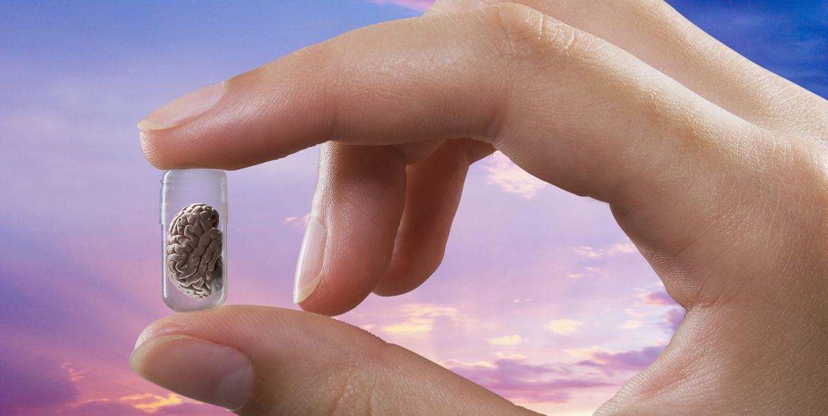
These gooey blobs of human brain matter could develop a type of intelligence we’ve never seen before.
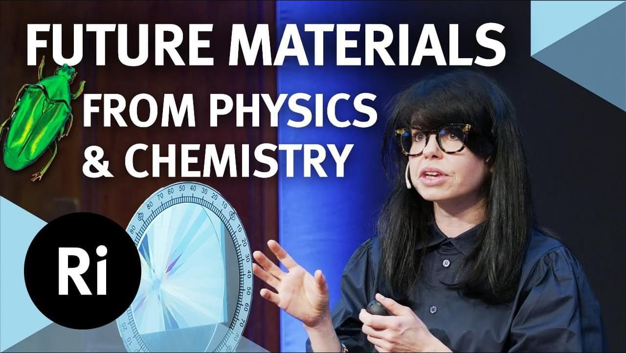
Jess Wade explains the concept of chirality, and how it might revolutionise technological innovation.
Join this channel to get access to perks:
https://www.youtube.com/channel/UCYeF244yNGuFefuFKqxIAXw/join.
Watch the Q&A here (exclusively for our Science Supporters): https://youtu.be/VlkHT-0zx9U
This lecture was recorded at the Ri on 14 June 2025.
Imagine if we could keep our mobile phones on full brightness all day, without worrying about draining our battery? Or if we could create a fuel cell that used sunlight to convert water into hydrogen and oxygen? Or if we could build a low-power sensor that could map out brain function?
Whether it’s optoelectronics, spintronics or quantum, the technologies of tomorrow are underpinned by advances in materials science and engineering. For example, chirality, a symmetry property of mirror-image systems that cannot be superimposed, can be used to control the spin of electrons and photons. Join functional materials scientist Jess Wade as she explores how advances in chemistry, physics and materials offer new opportunities in technological innovation.
–
Understanding how the human brain stores information and later uses it to complete various tasks has been a long-standing goal of neuroscience and psychology research. Past studies have identified different types of memory processes that have distinct roles and characteristics.
One of these, known as working memory, entails the storage and manipulation of important information for short periods of time, particularly information that is helpful for completing reasoning tasks or to make decisions in the short-term. Findings suggest that this temporary storage of information is associated with the continued and persistent firing of specific neurons in the brain.
Most past studies focusing on working memory processes relied on experimental tasks that require participants to prioritize and memorize all items they are presented with.

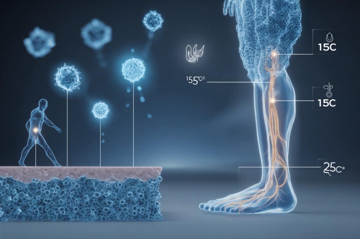
A personalized brain stimulation system powered by artificial intelligence (AI) that can safely enhance concentration from home has been developed by researchers from the University of Surrey, the University of Oxford and Cognitive Neurotechnology. Designed to adapt to individual characteristics, the system could help people improve focus during study, work, or other mentally demanding tasks.
Published in npj Digital Medicine, the study is based on a patented approach that uses non-invasive brain stimulation alongside adaptive AI to maximize its impact.
The technology uses transcranial random noise stimulation (tRNS)—a gentle and painless form of electrical brain stimulation—and an AI algorithm that learns to personalize stimulation based on individual features, including attention level and head size.
Childhood brain tumor survival depends on the type of tumor. Comparing survival rates across countries is difficult, because brain tumors aren’t recorded in the same way everywhere in Europe. A new study led by the Princess Máxima Center is helping to change that. For the first time, the research provides a clear and clinically relevant overview of survival outcomes for children with brain tumors.
Researchers at the Princess Máxima Center analyzed data from more than 30,000 children diagnosed with a brain tumor between 1998 and 2013. The data came from 80 cancer registries across 31 European countries. The study was published today in The Lancet Oncology.
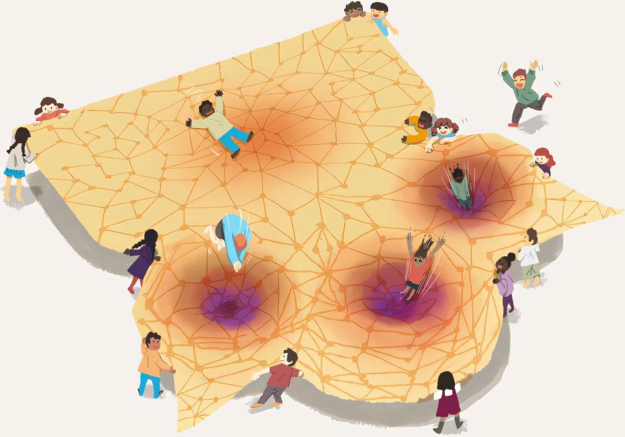
A new international study has introduced Curved Neural Networks—a new type of AI memory architecture inspired by ideas from geometry. The study shows that bending the “space” in which AI “thinks” can create explosive memory recall—an effect similar to a lightbulb moment in the human brain. The research opens new paths for brain-inspired computing, neuroscience, and even next-gen robotics, offering tools to better understand memory itself.
What if artificial intelligence could remember things not just well, but faster or more reliably? A new international study has introduced a novel type of AI memory —one that addresses the challenge not with more data, but with geometry.
A team of researchers from the Basque Center for Applied Mathematics (BCAM), Araya Inc., the University of Sussex, and Kyoto University has developed a new class of AI models called Curved Neural Networks.

In Alzheimer’s disease, proteins like amyloid beta form clumps, known as plaques, that damage the brain.
But in some people, immune cells called microglia break down these proteins before they can cause harm. This leads to fewer and smaller clumps—and much milder symptoms.
Researchers at UC San Francisco identified a molecular receptor that enables microglia to gobble up and digest amyloid beta plaques. The findings are published in the journal Neuron.
University of Kentucky Markey Cancer Center researchers have discovered a genetic biomarker that could help identify patients with glioblastoma most likely to benefit from the cancer drug bevacizumab.
The study, published in JCO Precision Oncology, found that brain tumors from patients treated with bevacizumab who lived longer were more likely to have a genetic change called CDK4 amplification. This suggests that testing for the molecular marker could help oncologists identify patients most likely to respond well to bevacizumab treatment.
“The findings could help oncologists make more informed treatment decisions for glioblastoma patients, potentially sparing those unlikely to benefit from unnecessary side effects while ensuring those who might respond get access to the drug,” said John Villano, M.D., Ph.D., the study’s lead author and professor in the UK College of Medicine.