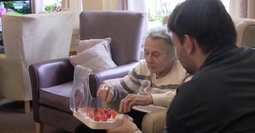
When London-based student Lewis Hornby noticed that his dementia-afflicted grandmother was having trouble staying hydrated, he came up with Jelly Drops, bite-sized pods of edible water made with gelling agents and electrolytes.
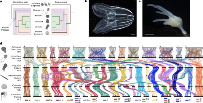
As sponges and ctenophores are such disparate animals13, the nature of the first diverging animal lineage has implications for the evolution of fundamental animal characteristics. Adult sponges are generally sessile filter-feeding organisms with body plans organized into reticulated water-filtration channels, structures built out of silica or calcium carbonate, and specialized cell types and tissues used for feeding, reproduction and self-defence, but they lack neuronal and muscle cells15. By contrast, ctenophores are gelatinous marine predators that move using eight longitudinal ‘comb rows’ of ciliary bundles16,17; they are superficially similar but unrelated to cnidarian medusae13,18 and possess multiple nerve nets19. Thus, whereas the sponge-sister scenario suggests a single origin of neurons on the ctenophore–parahoxozoan stem, the ctenophore-sister scenario implies either that either ancestral metazoan neurons were lost in the sponge lineage, or that there was convergent evolution of neurons in the ctenophore and parahoxozoan lineages3,6. Similar considerations apply to other metazoan cell types18, gene regulatory networks, animal development13,18 and other uniquely metazoan features.
Despite its importance for understanding animal evolution, the relative branching order of sponges, ctenophores and other animals has proven to be difficult to resolve2. The fossil record is largely silent on this issue as verified Precambrian sponge fossils are extremely rare20 and putative fossils of the soft-bodied ctenophores are difficult to interpret21. Morphological characters of living groups (for example, choanocytes of sponges) are not sufficient to resolve the question because true homology is difficult to assign, and such characters are easily lost or can arise convergently13,22. The ctenophore-sister hypothesis is supported by a pair of gene duplications shared by sponges, bilaterians, placozoans and cnidarians but not ctenophores23. Although sophisticated methods for sequence-based phylogenomics have been developed and applied to increasingly large molecular datasets, there is still considerable debate about the relative position of sponges and ctenophores as results are sensitive to how sequence evolution is modelled11, which taxa or sites are included24,25, and the effects of long-branch artifacts and nucleotide compositional variation26. New approaches are needed.
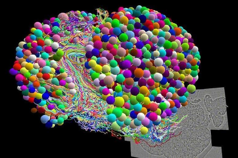
We reasoned that patterns of synteny, classically defined as chromosomal gene linkage without regard to gene order27, could provide a powerful tool for resolving the ctenophore-sister versus sponge-sister debate. Chromosomal patterns of gene linkage evolve slowly in many lineages12,28,29,30, probably because it is improbable for interchromosomal translocations to be fixed in populations with large effective population sizes28,31,32. Notably, some changes in synteny are effectively irreversible. For example, when two distinct ancestral synteny groups are combined onto a single chromosome by translocation, and subsequent intrachromosomal rearrangements mix these two groups of genes, it is very unlikely that the ancestral separated pattern will be restored by further rearrangement and fission, in the same sense that spontaneous reduction in entropy is improbable12. Such rare and irreversible changes are particularly useful for resolving challenging phylogenetic questions as they give rise to shared derived features that unambiguously unite all descendant lineages33,34,35. Deeply conserved syntenies observed between animals and their closest unicellular relatives12 suggest that outgroup comparisons could be used to infer ancestral metazoan states and polarize changes within animals to address the sponge-sister versus ctenophore-sister debate. Yet, chromosome-scale genome sequences of the unicellular or colonial eukaryotic outgroups closest to animals (choanoflagellates, filastereans and ichthyosporeans) have not been reported.
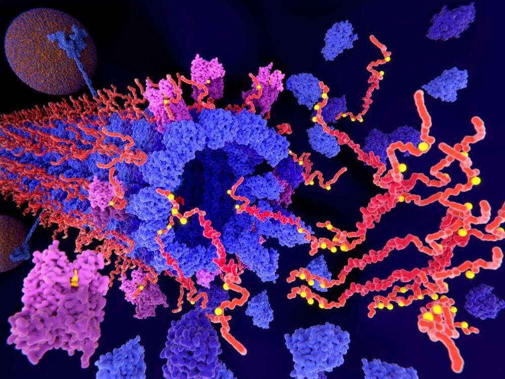
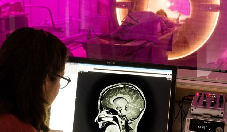
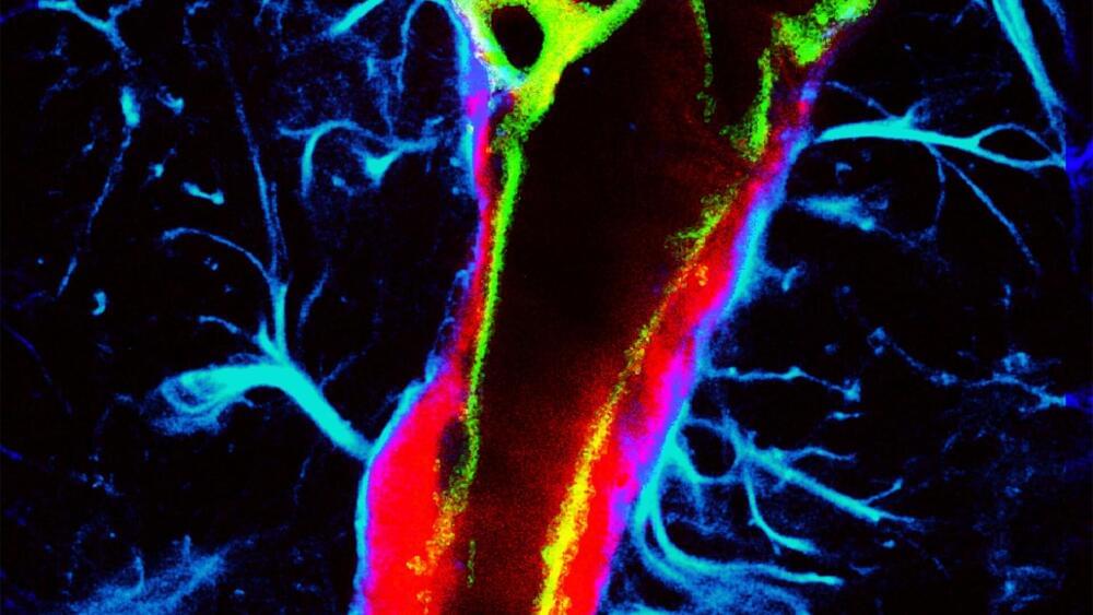
Like the lymphatic system in the body, the glymphatic system in the brain clears metabolic waste and distributes nutrients and other important compounds. Impairments in this system may contribute to brain diseases, such as neurodegenerative diseases and stroke.
A team of researchers in the McKelvey School of Engineering at Washington University in St. Louis has found a non-invasive and non-pharmaceutical method to influence glymphatic transport using focused ultrasound, opening the opportunity to use the method to further study brain diseases and brain function. Results of the work are published in the Proceedings of the National Academy of Sciences on May 15.
Hong Chen, associate professor of biomedical engineering in McKelvey Engineering and of neurological surgery in the School of Medicine, and her team, including Dezhuang (Summer) Ye, a postdoctoral research associate, and Si (Stacie) Chen, a former postdoctoral research associate, found the first direct evidence that focused ultrasound, combined with circulating microbubbles—a technique they call FUSMB—could mechanically enhance glymphatic transport in the mouse brain.
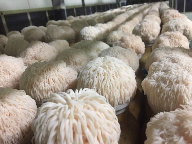
Lions mane the mushroom can actually stop alzheimers and dementia by boosting nerve growth 😗😁
Researchers from The University of Queensland have discovered the active compound from an edible mushroom that boosts nerve growth and enhances memory.
Professor Frederic Meunier from the Queensland Brain Institute said the team had identified new active compounds from the mushroom, Hericium erinaceus.
“Extracts from these so-called ‘lion’s mane’ mushrooms have been used in traditional medicine in Asian countries for centuries, but we wanted to scientifically determine their potential effect on brain cells,” Professor Meunier said.
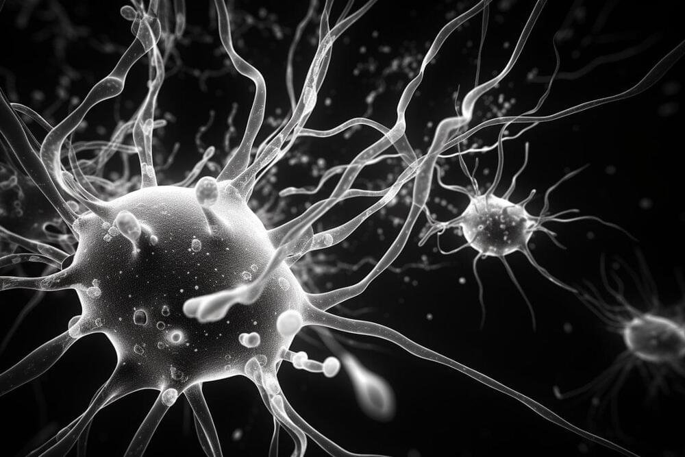
Summary: A new study discovered not all microglia are the same, challenging existing beliefs. A unique subset of these cells, the ARG1+microglia, important for proper cognitive functions, were identified in mice, with evidence suggesting a similar subset exists in humans.
Microglia lacking the protein ARG1 led to less exploratory behavior in mice, indicating cognitive deficits. These discoveries open exciting new possibilities for understanding brain diseases and developing novel therapies.
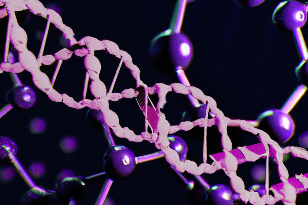
NewLimit, a company working towards the radical extension of human healthspan using epigenetic reprogramming has announced it has secured $40 million in Series A funding from prominent investors including Dimension, Founders Fund, and Kleiner Perkins.
This investment further bolsters the company’s belief that therapies to delay, halt or even reverse aging can be found through the exploration of epigenetic reprogramming. With a strong belief that their innovative approach can also address various age-related diseases, NewLimit aims to revolutionize the field of aging biology and pave the way for transformative advancements in healthcare.
Longevity. Technology: Epigenetic reprogramming is an emerging but exciting field of geroscience. It involves the identification of specific sets of transcription factors that can induce changes in gene expression and cellular behavior, effectively reversing or modifying the epigenetic markers associated with aging. This approach offers a unique opportunity to rejuvenate cells and tissues, potentially slowing down or even reversing the effects of aging and its related diseases. NewLimit says that while its products are designed to treat aging itself, the company also believes “these products could treat or prevent many diseases associated with aging, including fibrosis, infectious disease, and neurodegenerative disease.”
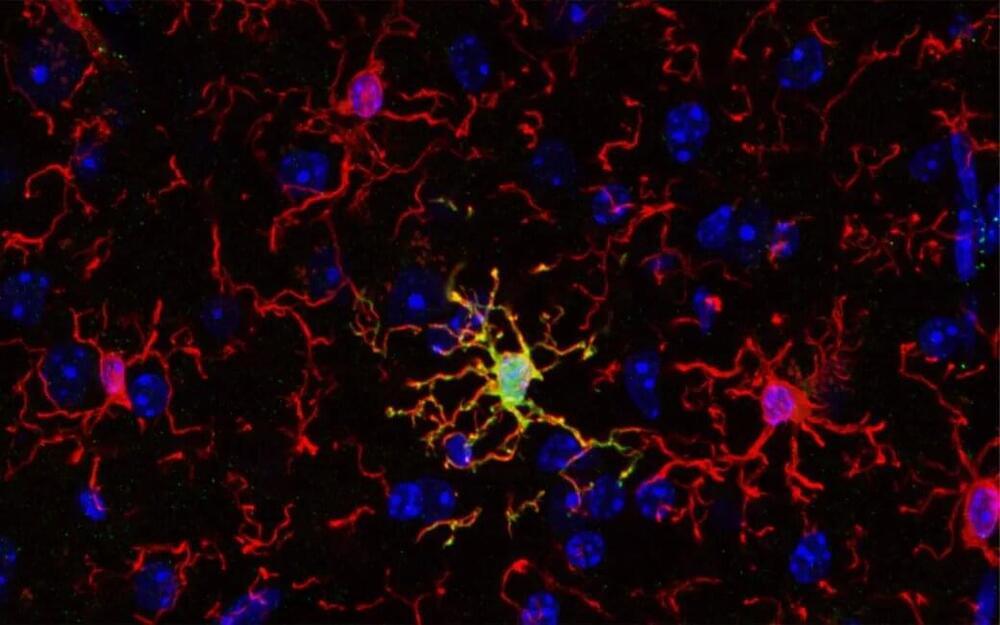
A recent study published in Nature Neuroscience indicates that, contrary to common belief, the immune cells of the brain, known as microglia, are not all the same. Researchers found that a unique microglial subset with unique features and function is important for establishing proper cognitive functions in mice. Evidence for such microglial subsets exists also for the human brain, opening exciting new possibilities for novel therapies.
An international collaboration led by researchers from University of Helsinki, Karolinska Institutet and University of Seville characterized ARG1+ microglia, a subset of microglial cells, that produces the enzyme called arginase-1 (ARG1). Using advanced imaging techniques, the team found that ARG1+ microglia are abundant during development and less prevalent in adult animals. Strikingly, these ARG1+ microglia are located in specific brain areas important for cognitive functions such as learning, thinking and memory.
“Cognition and memory are crucial components of what makes us human, and microglia are necessary for proper brain development and function. Cognitive decline is a common feature of neurodegenerative and psychiatric conditions like Alzheimer’s and Parkinson’s disease, schizophrenia and depression,” says Dr. Vassilis Stratoulias, senior researcher at the University of Helsinki and lead author of the study.
The touch of another person may increase levels of the “feel-good” hormone oxytocin. But the context really matters. The situation impacts oxytocin levels not only in the moment, but also later, as is shown by researchers at Linköping University and the University of Skövde in Sweden. Their study has been published in the journal eLife.
An embrace from a parent, a warm hand on your shoulder or a caress from a romantic partner are examples of how touch can strengthen social bonds between people and influence emotions. But although touch and the sense of touch have a very important function, knowledge of how this actually works is still lacking.
Studies in animals have shown that the hormone oxytocin is linked to touch and social bonding. However, many questions remain unanswered when it comes to oxytocin’s role in human social interactions and how this hormone can influence and be influenced by the brain. To study this more closely, researchers have examined what happens in the body when we feel a soft touch.