A 25-year-old wager is settled as a new generation of scientists takes on competing theories of consciousness.
When humans make decisions, such as picking what to eat from a menu, what jumper to buy at a store, what political candidate to vote for, and so on, they might be more or less confident with their choice. If we are less confident and thus experience greater uncertainty in relation to their choice, our choices also tend to be less consistent, meaning that we will be more likely to change our mind before reaching a final decision.
While neuroscientists have been exploring the neural underpinnings decision-making for decades, many questions are still unanswered. For instance, how neural network computations support decision-making under varying levels of certainty remain poorly understood.
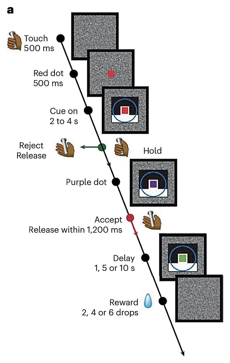
Researchers at the National Institute of Mental Health in Bethesda, Maryland recently carried out a study on rhesus monkeys aimed at better understanding the neural network dynamics associated with decision confidence. Their paper, published in Nature Neuroscience, offers evidence that energy landscapes in the prefrontal cortex can predict the consistency of choices made by monkeys, which is in turn a sign of the animals’ confidence in their decisions.
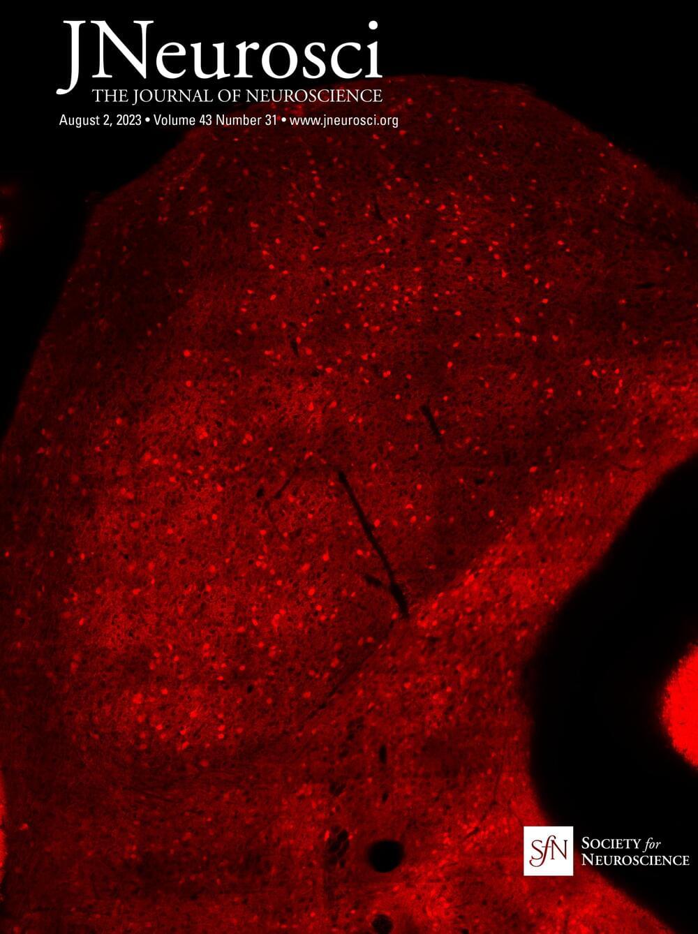
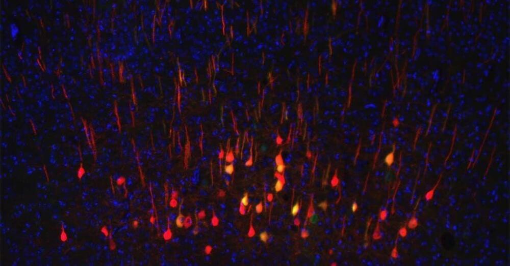
The parabrachial nuclear complex (PBN) is a nexus for aversion and for the sensory and affective components of pain perception. We have previously shown that during chronic pain PBN neurons in anesthetized rodents have amplified activity. We report a method to record from PBN neurons of behaving, head-restrained mice while applying reproducible noxious stimuli. We find that both spontaneous and evoked activity are higher in awake animals compared with urethane anesthetized mice. Fiber photometry of calcium responses from calcitonin-gene-related peptide–expressing PBN neurons demonstrates that these neurons respond to noxious stimuli. In both males and females with neuropathic or inflammatory pain, responses of PBN neurons remain amplified for at least 5 weeks, in parallel with increased pain metrics. We also show that PBN neurons can be rapidly conditioned to respond to innocuous stimuli after pairing with noxious stimuli. Finally, we demonstrate that changes in PBN neuronal activity are correlated with changes in arousal, measured as changes in pupil area.
SIGNIFICANCE STATEMENT The parabrachial complex is a nexus of aversion, including pain. We report a method to record from parabrachial nucleus neurons of behaving mice while applying reproducible noxious stimuli. This allowed us to track parabrachial activity over time in animals with neuropathic or inflammatory pain. It also allowed us to show that the activity of these neurons correlates with arousal states and that these neurons can be conditioned to respond to innocuous stimuli.

How basic income works.
Over the course of a year, the Denver Basic Income Project gives participants cash payments of varying amounts. Many participants, some of whom were living on the streets a few months before enrolling in the program, reported feeling safer, happier, and less anxious with better living arrangements.
The Denver Basic Income Project began in 2021 and was granted a $2 million contribution from the city. Researchers at the University of Denver’s Center on Housing and Homelessness Research found most of those who received money from the program were significantly better off six months in.
Dia Broncucia and Justin Searls bought a car, rented an apartment, and improved their mental health after receiving monthly basic-income payments.
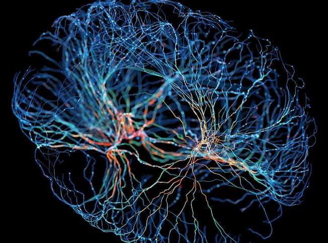
The human brain is made up of two kinds of matter: the nerve cell bodies (gray matter), which process sensation, control voluntary movement, and enable speech, learning and cognition, and the axons (white matter), which connect cells to each other and project to the rest of the body.
Historically, scientists have concentrated on the gray matter of the cortex, figuring that’s where the action is, while ignoring white matter, even though it makes up half the brain. Researchers at Vanderbilt University are out to change that.
For several years, John Gore, Ph.D., director of the Vanderbilt University Institute of Imaging Science, and his colleagues have used functional magnetic resonance imaging (fMRI) to detect blood oxygenation-level dependent (BOLD) signals, a key marker of brain activity, in white matter.
Scientists at Kyushu University report that turning brain immune cells into neurons restores brain function after stroke-like injury in mice. Their findings suggest that replenishing neurons from immune cells could be a potential avenue for treating stroke in humans.
The findings are published in PNAS in an article titled, “Direct neuronal conversion of microglia/macrophages reinstates neurological function after stroke.”
“Although generating new neurons in the ischemic injured brain would be an ideal approach to replenish the lost neurons for repairing the damage, the adult mammalian brain retains only limited neurogenic capability,” wrote the scientists. “Here, we show that direct conversion of microglia/macrophages into neurons in the brain has great potential as a therapeutic strategy for ischemic brain injury.”
A recent study in Psychiatry Research: Neuroimaging revealed that individuals with high depression scores show increased activity in frontal brain regions during visuospatial memory tasks, despite similar behavioral performance to those with low depression scores. Researchers concluded that the heightened brain activity might represent a compensatory effort…
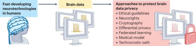
Neurotechnology will improve our lives in many ways. However, to sustain a world where our neurobiological data (in some cases perhaps including our innermost thoughts and feelings) remains properly secure, we must invest in both policy and technology that prevents bad actors from stealing private information or even directly manipulating people’s brains. We don’t want the very real possibility of ‘telepathy’ and ‘mind control’ to harm people and society. So, let’s start laying the groundwork now to ensure the best possible neurotech future! #neurotech #future #policy #neuroscience
We provide a Perspective highlighting the significant ethical implications of the use of fast-developing neurotechnologies in humans, as well as the regulatory frameworks and guidelines needed to protect neurodata and mental privacy.
Neurons, the main cells that make up our brain and spinal cord, are among the slowest cells to regenerate after an injury, and many neurons fail to regenerate entirely. While scientists have made progress in understanding neuronal regeneration, it remains unknown why some neurons regenerate and others do not.
Using single-cell RNA sequencing, a method that determines which genes are activated in individual cells, researchers from University of California San Diego School of Medicine have identified a new biomarker that can be used to predict whether or not neurons will regenerate after an injury. Testing their discovery in mice, they found that the biomarker was consistently reliable in… More.
Researchers from University of California San Diego have identified a new biomarker that can predict whether or not neurons will regenerate after an injury. The findings could help scientists develop regenerative therapies for spinal cord injuries and other neurological conditions.