Stem cell biologist Helen Blau of Stanford University School of Medicine and colleagues previously found that blocking 15-PGDH in old mice restored their withered muscles and improved their strength after a month of treatment. On the flip side, young mice lost muscle and became weaker after their levels of this enzyme were increased for a month.
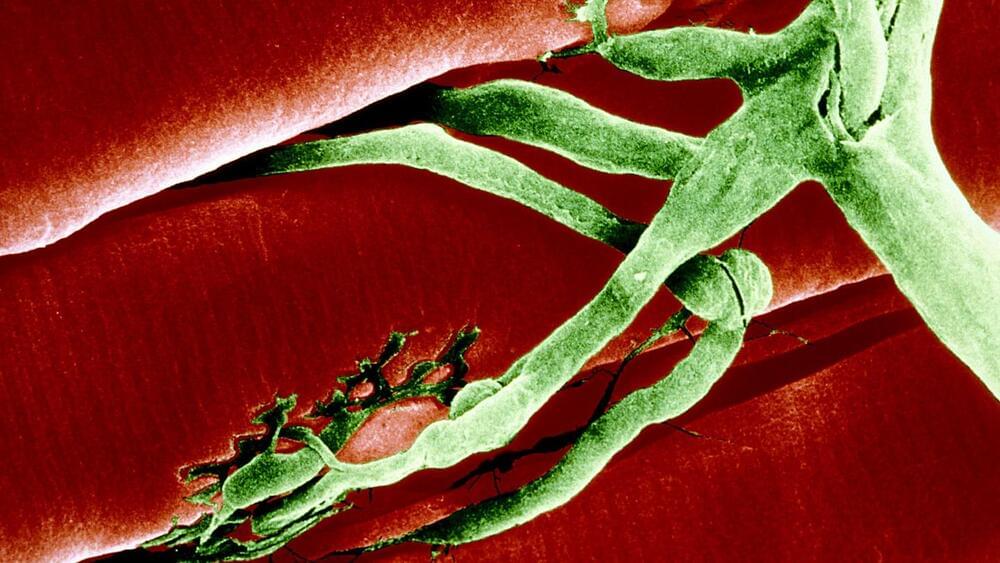
Blau’s team has now found that 15-PGDH accumulates in the muscles of old mice as the connections that allow communication between muscles and nerves are lost, another consequence of aging. Treating old mice for one month with a drug that inhibits 15-PGDH restored these connections, called synapses, between muscle fibers and motor nerve cells, and boosted the animals’ strength, the team reports in the Oct. 11 Science Translational Medicine. Those synapses are how the brain directs muscles to move.
The findings suggest that blocking the gerozyme 15-PGDH may be a way to help recover strength that has diminished due to nerve injuries, motor nerve cell diseases or aging.