However, underlying this scientific skepticism was also an ideological shift. Reductionism can be thought of as the antithesis or critique of the concepts of a premodern worldview. The rejection of the self was motivated by a hidden agenda to rid science of any ideas that remotely felt supernatural or religious. Since the self seemed intertwined with the idea of a soul, scientific pushback on ideological grounds was inevitable, and from that point on, findings from neuroscience and psychology were interpreted through a reductionist lens. The fact that scientists could not identify a localized region that precisely corresponded to the self seemed to verify the belief that it is an “illusion,” though to most people that statement has little meaning, if any.
This reductionist ideology recently found an ally in what is called “nondual” Eastern philosophy. According to this quasi-mystical doctrine, embracing the idea that we aren’t our thoughts or ego can lead to a more compassionate world — one free of self-blame and blame toward others. If none of us are in control of our actions or thoughts, then punishment is pointless and immoral. By not placing undue importance on the self, individuals might find themselves more attuned to the interconnected nature of existence, shifting toward a holistic worldview where “we’re all in this together.”
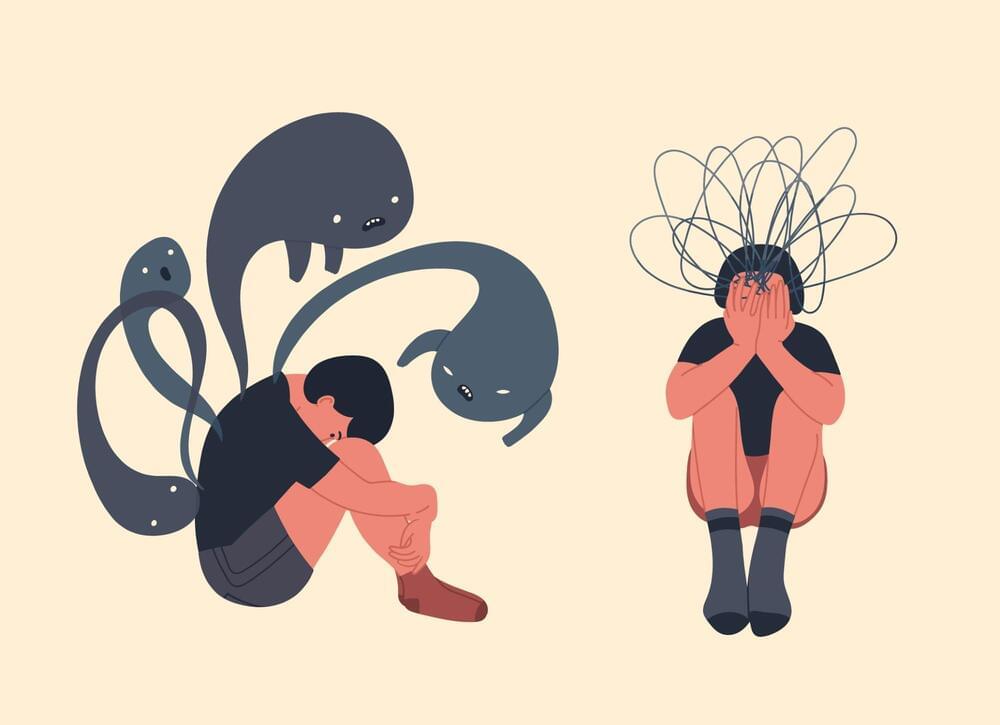
However, there’s a dark side to this denial of the self, and it’s extremely troubling to those who think about this stuff deeply. If we have no self and no control over our thoughts and actions, then we are slaves to a billiard ball universe, trapped in a nihilistic nightmare in which we cannot change our fate or the fate of humanity. For those who take the hardline reductionist stance seriously, this can lead to cognitive dissonance, and in rarer cases, crippling depression or psychosis.