We construct our experiences.
Category: neuroscience – Page 675
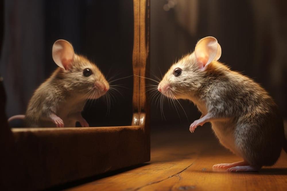
Mirror Insight: Mice Show Glimpses of Self-Recognition
Summary: Mice display behavior akin to self-recognition when viewing their reflections in mirrors. This behavior emerges under specific conditions: familiarity with mirrors, socialization with similar-looking mice, and visible markings on their fur.
The study also identifies a subset of neurons in the hippocampus that are crucial for this self-recognition-like behavior. These findings provide valuable insights into the neural mechanisms behind self-recognition, a previously enigmatic aspect of neurobehavioral research.
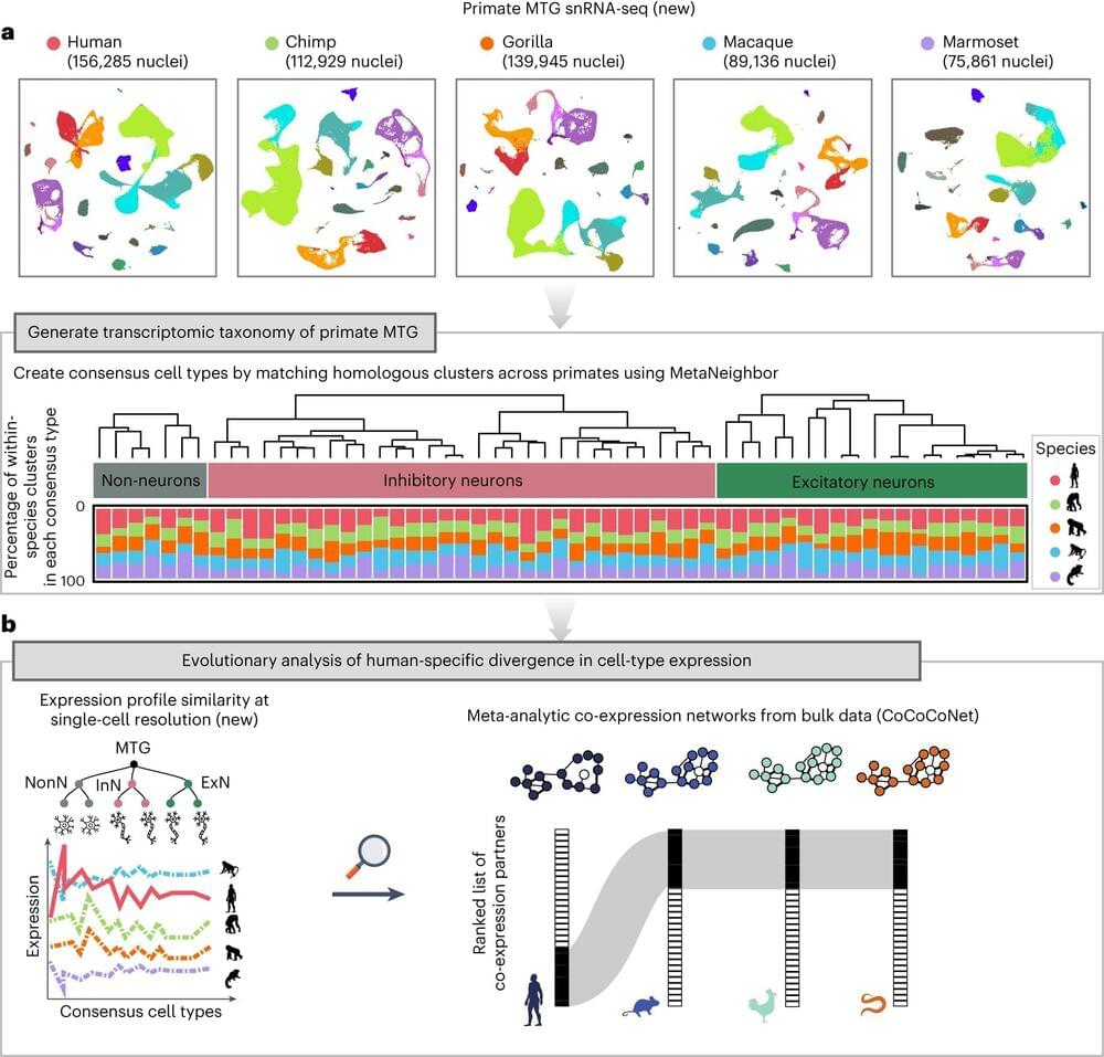
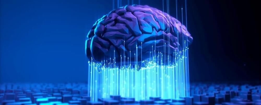
Study reveals genes that set humans apart from other primates in cognitive ability
An international team led by researchers at the University of Toronto has uncovered over 100 genes that are common to primate brains but have undergone evolutionary divergence only in humans—and which could be a source of our unique cognitive ability.
The researchers, led by Associate Professor Jesse Gillis from the Donnelly Center for Cellular and Biomolecular Research and the department of physiology at U of T’s Temerty Faculty of Medicine, found the genes are expressed differently in the brains of humans compared to four of our relatives—chimpanzees, gorillas, macaques and marmosets.
The findings, published in Nature Ecology & Evolution, suggest that reduced selective pressure, or tolerance to loss-of-function mutations, may have allowed the genes to take on higher-level cognitive capacity. The study is part of the Human Cell Atlas, a global initiative to map all human cells to better understand health and disease.
#BuildFor2030: Empowering an inclusive and accessible world
📸 Watch this video on Facebook https://www.facebook.com/share/v/dv8r6G2mywg5NthT/?mibextid=qi2Omg
Accessibility empowers innovation for everyone.
Technology can empower the 1+ billion people living with disabilities to achieve more, and the benefit is for us all. Solutions built with inclusive design and accessibility top of mind can help improve our experience, productivity, and engagement.
We’re proud of Microsoft partners empowering a more accessible world. These partners are driving innovation in assistive technology, enabling more inclusive workplaces, and increasing disability inclusion and support for mental health needs in their communities. Join us in supporting partners building for a more inclusive and accessible world.

AI Revolutionizes Neuron Tracking in Moving Animals
Summary: Researchers developed an AI-based method to track neurons in moving and deforming animals, a significant advancement in neuroscience research. This convolutional neural network (CNN) method overcomes the challenge of tracking brain activity in organisms like worms, whose bodies constantly change shape.
By employing ‘targeted augmentation’, the AI significantly reduces the need for manual image annotation, streamlining the neuron identification process. Tested on the roundworm Caenorhabditis elegans, this technology has not only increased analysis efficiency but also deepened insights into complex neuronal behaviors.

Earth on verge of five catastrophic climate tipping points, scientists warn
Recently, economists and behavioral scientists have studied the pattern of human well-being over the lifespan. In dozens of countries, and for a large range of well-being measures, including happiness and mental health, well-being is high in youth, falls to a nadir in midlife, and rises again in old age. The reasons for this U-shape are still unclear. Present theories emphasize sociological and economic forces. In this study we show that a similar U-shape exists in 508 great apes (two samples of chimpanzees and one sample of orangutans) whose well-being was assessed by raters familiar with the individual apes. This U-shaped pattern or “midlife crisis” emerges with or without use of parametric methods. Our results imply that human well-being’s curved shape is not uniquely human and that, although it may be partly explained by aspects of human life and society, its origins may lie partly in the biology we share with great apes. These findings have implications across scientific and social-scientific disciplines, and may help to identify ways of enhancing human and ape well-being.

Evidence for a midlife crisis in great apes consistent with the U-shape in human well-being
Recently, economists and behavioral scientists have studied the pattern of human well-being over the lifespan. In dozens of countries, and for a large range of well-being measures, including happiness and mental health, well-being is high in youth, falls to a nadir in midlife, and rises again in old age. The reasons for this U-shape are still unclear. Present theories emphasize sociological and economic forces. In this study we show that a similar U-shape exists in 508 great apes (two samples of chimpanzees and one sample of orangutans) whose well-being was assessed by raters familiar with the individual apes. This U-shaped pattern or “midlife crisis” emerges with or without use of parametric methods. Our results imply that human well-being’s curved shape is not uniquely human and that, although it may be partly explained by aspects of human life and society, its origins may lie partly in the biology we share with great apes. These findings have implications across scientific and social-scientific disciplines, and may help to identify ways of enhancing human and ape well-being.
Uploading Your Mind to a Computer Will Require 3 Crucial Things
Imagine brain scanning technology improves greatly in the coming decades, to the point that we can observe how each individual neuron talks to other neurons.
Then, imagine we can record all this information to create a simulation of someone’s brain on a computer.
This is the concept behind mind uploading – the idea that we may one day be able to transition a person from their biological body to a synthetic hardware.