Toggle light / dark theme

Scientists cast light on the brain’s social cells

Picture yourself hovering over an alien city with billions of blinking lights of thousands of types, with the task of figuring out which ones are connected, which way the electricity flows and how that translates into nightlife. Welcome to the deep brain.

Even in an era rapidly becoming known as the heyday of neuroscience, tracing the biochemical signaling among billions of neurons deep in the brain has remained elusive and baffling.

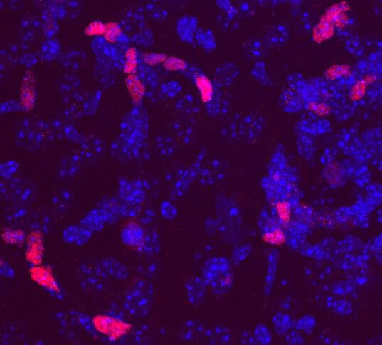
A team of Stanford University researchers managed to map out one such connection, buried inside the brain of a living, moving mammal as they manipulated its behavior. The feat offers an unprecedented close-up of the genesis of on a cellular level, and could offer insights into psychiatric puzzles such as autism, depression and anxiety.
Study charts development of emotional control in teens

In the midst of all the apparent tumult, intense emotion, and occasional reckless behavior characterizing the teenage years, the brain is, in fact, evolving and developing the neural circuits needed to keep emotions in check. Research in the June 8, 2016 issue of The Journal of Neuroscience describes how the ability to control emotions moves from one brain area to another as teens mature into adults, offering an opportunity to understand how disorders related to emotional control emerge.

“Our study opens the way for a better understanding of the neurobiology behind adolescent behavior in emotionally arousing situations,” said study author Anna Tyborowska of Radboud University Nijmegen in the Netherlands. “The findings could also have important clinical implications [as] many psychiatric disorders emerge during adolescence and are characterized by problems with emotional action control.”

Previous research links the spike in sensation-seeking and impulsive behavior during adolescence to the delayed maturation of the , a region of the involved in reasoning, planning, and decision-making. Study authors Inge Volman, Ivan Toni, and Karin Roelofs previously demonstrated the importance of the anterior prefrontal cortex in emotional control in adults. However, it has not been clear whether and how the delayed development of the prefrontal cortex affects emotional control during adolescence.

New computational tool could help optimize treatment of Alzheimer’s disease

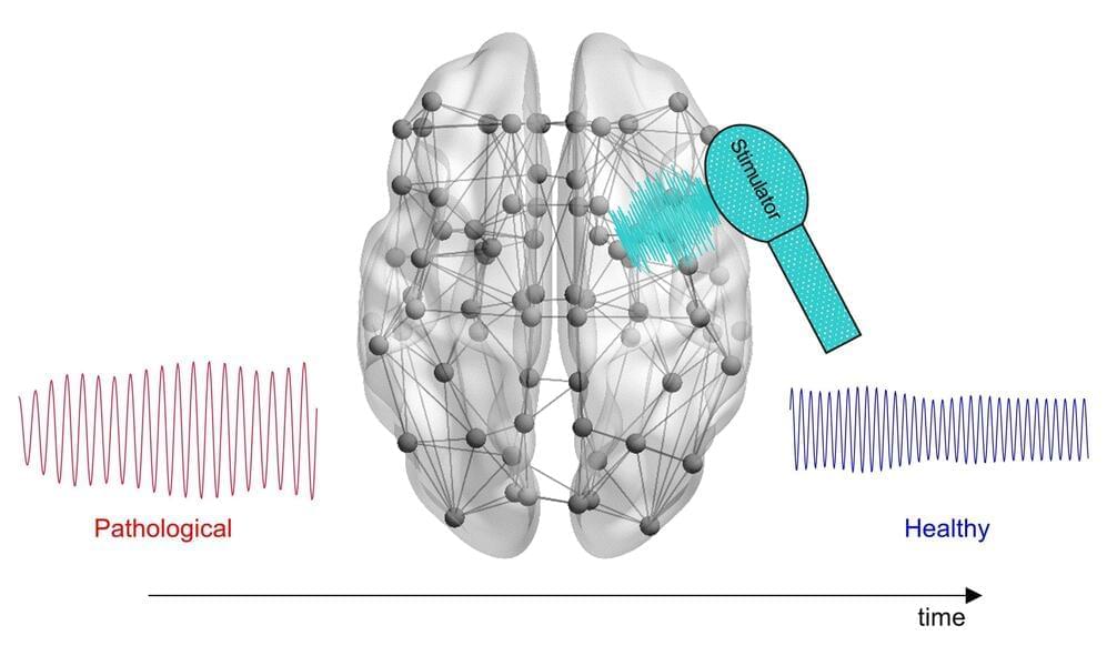
Scientists have developed a novel computational approach that incorporates individual patients’ brain activity to calculate optimal, personalized brain stimulation treatment for Alzheimer’s disease. Lazaro Sanchez-Rodriguez of the University of Calgary, Canada, and colleagues present their new framework in PLOS Computational Biology.

Electrical stimulation of certain parts of the could help promote healthy activity in neural circuits impaired by Alzheimer’s disease, a neurodegenerative condition. This experimental treatment has shown some promise in . However, all patients currently receive identical treatment protocols, potentially leading to different outcomes according to individual variations in brain signaling.

To investigate the possibility of personalized brain stimulation, Sanchez-Rodriguez and colleagues took a theoretical approach. They built a computational tool that incorporates patients’ MRI scans and physiological brain signaling measurements to calculate optimal brain stimulation signals, with the goal of delivering efficient, effective personalized treatment.

Low strength brain stimulation may be effective for depression

Brain stimulation treatments, like electroconvulsive therapy (ECT) and transcranial magnetic stimulation (TMS), are often effective for the treatment of depression. Like antidepressant medications, however, they typically have a delayed onset. For example, a patient may receive several weeks of regular ECT treatments before a full response is achieved.

Thus, there is an impetus to develop antidepressant treatments that act to rapidly improve mood.

Low field magnetic stimulation (LFMS) is one such potential new with rapid mood-elevating effects, as reported by researchers at Harvard Medical School and Weill Cornell Medical College.

Epileptic seizures and depression may share a common genetic cause, study suggests

From the time of Hippocrates, physicians have suspected a link between epilepsy and depression. Now, for the first time, scientists at Rutgers University-New Brunswick and Columbia University have found evidence that seizures and mood disorders such as depression may share the same genetic cause in some people with epilepsy, which may lead to better screening and treatment to improve patients’ quality of life.

The scientists studied dozens of unusual families with multiple relatives who had epilepsy, and compared the family members’ of with that of the U.S. population.

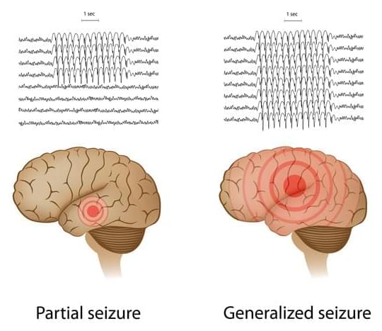
They found an increased incidence of mood disorders in persons who suffer from a type of the condition called focal epilepsy, in which begin in just one part of the brain. But mood disorders were not increased in people with generalized epilepsy, in which seizures start on both sides of the brain.

Overnight brain stimulation improves memory

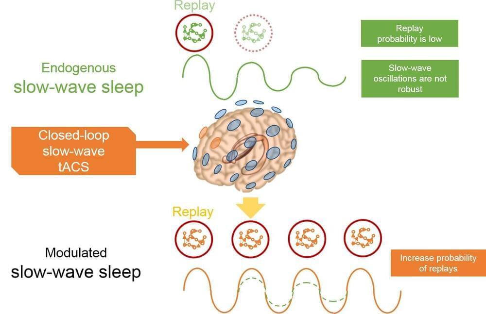
New research in humans demonstrates the potential to improve memory with a non-invasive brain stimulation technique delivered during sleep. The results, published in JNeurosci, come from a project funded by the United States Department of Defense that aims to better understand the process of memory consolidation, which could translate into improved memory function in both healthy and patient populations.

The transfer of memories from the hippocampus to the neocortex for long-term storage is thought to be enabled by synchronization of these parts of the brain during sleep. Nicholas Ketz, Praveen Pilly, and colleagues at University of New Mexico sought to enhance this natural process of overnight reactivation or neural replay to improve with a closed-loop transcranial alternating current stimulation system matching the phase and frequency of ongoing slow-wave oscillations during sleep.

Participants were trained and tested on a realistic visual discrimination task in which they had to detect potentially threatening hidden objects and people such as explosive devices and enemy snipers. The researchers found that when participants received stimulation during overnight visits to their sleep laboratory, they showed improved performance in detecting targets in similar but novel situations the next day compared to when they did not receive the stimulation, suggesting an integration of recent experience into a more robust and general memory. Overnight memory changes correlated with stimulation-induced neural changes, which could be used to optimize stimulation in future applications. These findings provide a method for enhancing without disturbing sleep.

A Common Link Between Several Neurodegenerative Diseases Might Finally Be Identified

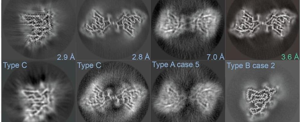
There’s a hallmark of incurable neurodegenerative diseases – misfolded proteins that clump together to form sticky plaques or tangles called fibrils.

Now, new research has discovered that a protein normally tasked with clearing cells of molecular debris might be a common feature of a cluster of common and rare neurodegenerative diseases, including two distinct forms of dementia.

The finding was “both unexpected and surprising” and “raises many intriguing questions”, according to the team behind the study, who made 3D-reconstructions of a twisted protein they found in “copious amounts” in some brain tissue samples.

Cool Inventions That Will Kill Your Boredom

BRAIN TIME ► https://goo.gl/tTWgH2

1. Parajet Xplorer Flying Car.

https://www.youtube.com/channel/UCTuvhzGL2APrK4jZ2ARX3pw.

2. HoverKart (Preview)
https://www.kickstarter.com/projects/hoverpowered/hoverkart-…-an-awesom.

3. Widescape WS250

https://www.youtube.com/channel/UCpVgPF1zITb2RjnO91RnPVw.

4. Overboat https://www.youtube.com/watch?v=mpOdIM8D9gc.
https://www.youtube.com/watch?v=pF1qoU7p1-k.
https://www.youtube.com/watch?v=hdqKzdhZvts.
https://www.youtube.com/watch?v=Trjr_bpMr0A
https://www.youtube.com/channel/UCJw0AF5K-k9NHGkdjEu7wjQ

5. All-Pro Passer.

Single protein prompts mature brain cells to regenerate multiple cell types

A single protein can reverse the developmental clock on adult brain cells called astrocytes, morphing them into stem-like cells that produce neurons and other cell types, UT Southwestern researchers report in a PNAS study. The findings might someday lead to a way to regenerate brain tissue after disease or injury.

“We’re showing that it may be possible to reprogram the fate of this subset of brain , giving them the potential to rebuild the damaged brain,” said study leader and co-corresponding author Chun-Li Zhang, Ph.D., Professor of Molecular Biology and an Investigator in the Peter O’Donnell Jr. Brain Institute.

During development, mammalian stem cells readily proliferate to produce neurons throughout the brain and cells—called glia—that help support them. Glia help maintain optimal brain function by performing essential jobs like cleaning up waste and insulating nerve fibers. However, the mature brain largely loses that stem cell capacity. Only two small regenerative zones, or niches, remain in the adult brain, Dr. Zhang explained, leaving it with extremely limited capacity to heal itself following injury or disease.

New Discoveries Reveal Information’s Flow in the Brain

Could these connections formulate our consciousness? Is consciousness information flowing throughout the brain?

The Neuro-Network.

See more.


A long-standing research collaboration is simultaneously recording populations of neurons across multiple brain areas in the visual system and utilizing novel statistical methods to observe neural activity patterns being conveyed between the areas.