Discount Links: Epigenetic, Telomere Testing: https://trudiagnostic.com/?irclickid=U-s3Ii2r7xyIU-LSYLyQdQ6…irgwc=1Use Code: CONQUERAGINGNAD+…
Category: neuroscience – Page 623
How Does the Brain Make Decisions? Harvard Scientists Shed New Light
Researchers have uncovered new insights into the way brain cells, or neurons, interact when making a decision, and how the links between these neurons could reinforce a decision.
The study — conducted in mice and led by neuroscientists at Harvard Medical School — is the first to combine structural, functional, and behavioral analyses to explore how neuron-to-neuron connections support decision-making.
The findings were recently published in the journal Nature.
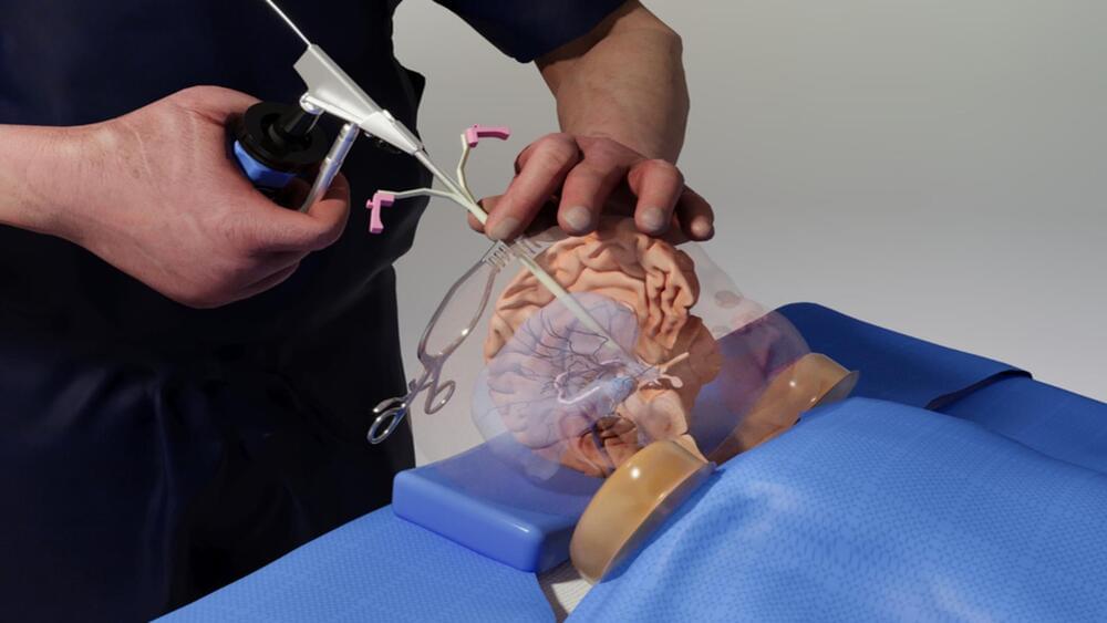
In a First, Organoid Model Resembles All Three Sections of Embryonic Brain and Spinal Cord
The first organized stem cell culture model that resembles all three sections of the embryonic brain and spinal cord, and produces a full model of the early stages of the human central nervous system, has been developed by a team of engineers and biologists at the University of Michigan(U-M), the Weizmann Institute of Science, and the University of Pennsylvania (UPenn).
“Models like this will open doors for fundamental research to understand early development of the human central nervous system and how it could go wrong in different disorders,” said Jianping Fu, PhD, professor of mechanical engineering at University of Michigan.
This work is published in Nature in the paper, “A Patterned Human Neural Tube Model Using Microfluidic Gradients.”
David Eagleman, investigator of the secrets of our minds
Nothing of the mind is foreign to David Eagleman, neuroscientist, technologist, entrepreneur and one of the most interesting scientific writers of our time. Born in New Mexico 52 years ago, he now researches cerebral plasticity, synesthesia, perception of time and what he called neurolaw, the intersection of the brain’s knowledge and its legal implications. His 2011 book Incognito: The Secret Lives of the Brain has been translated into 28 languages, and he returned to publishing with Livewired: The Inside Story of the Ever-Changing Brain, which focuses on a fundamental idea for today’s neuroscience: that the brain is constantly changing to be able to adapt to experience and learning. The science he brings to us isn’t merely top-notch, but firsthand, and his brilliant, crystal-clear writing — a perfect reflection of his mind — turns one of the most complex subjects of modern-day research into food for thought for the interested reader. We spoke with him in California by videoconference, the first interview that he’s given to a Spanish publication in a decade.
Could a newborn brain learn to live in a five-dimensional word? “We don’t actually know which things are pre-programmed and how much is experiential in our brains,” he replies. “If you could raise a baby in a five-dimensional world, which, of course, is unethical to do as an experiment, you might find that it’s perfectly able to function in that world. The general story of brain plasticity is that everything is more surprising than we thought, in terms of the brain’s ability to learn whatever world it drops into.”
Eagleman pulls out a sizable bowl of salad from somewhere, scoops a forkful into his mouth and continues his argument: “The five-dimensional world is hypothetical, but what we do see, of course, is that babies dropping into very different cultures around the planet, whether that’s a hyper-religious culture or an atheist culture, whether it’s a culture that lives on agriculture or a culture that is super technically advanced like here in Silicon Valley, the brain has no problem adjusting. My kids, when they were very young, could operate an iPad or cell phone just as easily as somebody growing up in a different place would operate farming equipment. So, we do know that brains are extremely flexible.”

Ableism Puts Neurodivergent Students at a Disadvantage
Physics has a diversity problem: those with identities outside of the majority “able-bodied, white, cis, and male” face significant barriers to entry. While efforts in the US to level the playing field are beginning to show success, studies continue to find that minority physicists will likely experience some form of bigotry, bias, or barrier during their career that will hamper their chances of success. These inequities and biases range from skewed course structures that favor specific learning styles (see Research News: Restructuring Classes Can Level the Playing Field) to systemic prejudices that hinder some groups from gaining grants (see News Feature: Systemic Racism Reflected in Grant Allocations, Researchers Argue) to unconscious biases that lead to the significant undercitation of minority physicists compared to their white, male counterparts (see News Feature: The Uneven Spread of Citations). All these factors can have serious career consequences, with negative experiences being a key factor driving people to leave the field.
One lesser-studied aspect of identity and how it impacts a person’s experience in physics is neurodivergence—a nonmedical umbrella term used to describe people whose brains process information in way that is different to what is considered normal. Now Geraldine Cochran of Ohio State University and Liam McDermott and Nazeer Mosley, both of Rutgers University, New Jersey, have developed a framework for interpreting the experiences of this group of people [1]. An initial analysis of interviews with three neurodivergent physicists shows that, while this group reports little outright discrimination or violence, structural ableism negatively impacted their time as students. “There are more neurodivergent people entering college than ever before,” McDermott says. “But their needs regularly get overlooked.”
A person who identifies as neurodivergent may have a neurological disorder, such as autism or Tourette’s syndrome; they may have a learning disability, such as dyslexia (which affects language processing) or dyscalculia (which affects number processing); or they could have a mental illness, such as depression or anxiety. For their study, Cochran, McDermott, and Mosley interviewed three physicists who identified as being neurodivergent and who pursued nonacademic careers after completing their undergraduate degrees. All three identified as having attention-deficit hyperactivity disorder (ADHD) and anxiety. Sky (the interviewees were all given pseudonyms) also has depression, Catalina has depression and dyslexia, and Henry has epilepsy. The interviews covered the trio’s undergraduate experiences. Cochran, McDermott, and Mosley then analyzed the trio’s answers using their newly developed “Critical Disability Physics Identity” framework.
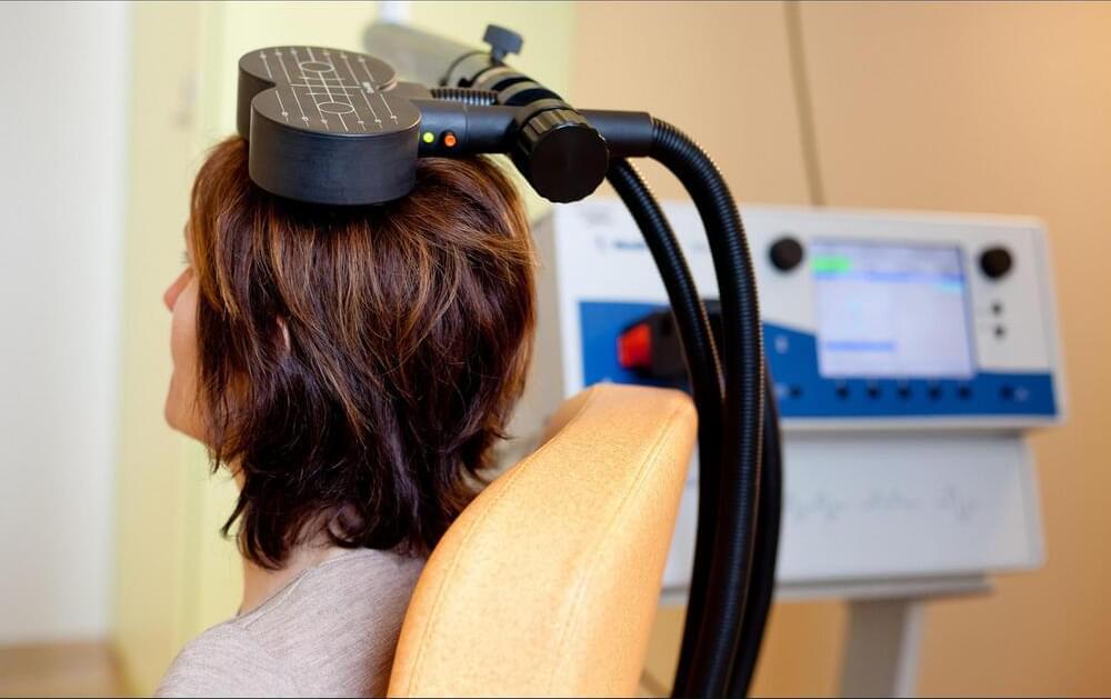
Brain stimulation poised to move from last resort to frontline treatment
Get alerts for new articles, or get an alert when an article is cited.
Even so, proponents say that TMS and other noninvasive brain-stimulation methods—which include updated forms of electroconvulsive therapy (ECT) and transcranial direct-current stimulation—have yet to achieve their full potential, both as research tools and as clinical treatments for a range of neurological conditions. To get there, researchers want to fully understand the biological mechanisms behind these techniques, along with finding more rigorous ways to test them in the lab, all with a view toward making treatments more tailored and reliably successful. With its demonstrated benefits and lack of serious side effects, Colleen Loo, a neurostimulation pioneer at the University of New South Wales, says, “there’s no reason TMS can’t be used as a frontline treatment” for major depression.