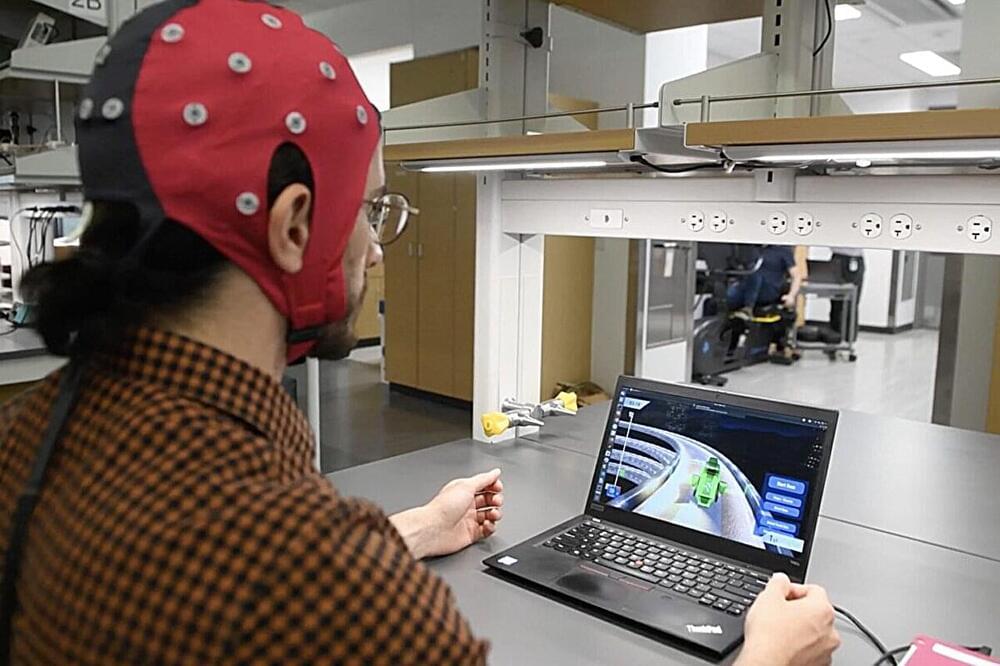
Imagine playing a racing game like Mario Kart, using only your brain to execute the complex series of turns in a lap.

A new study indicates a link between sleep apnea and increased memory or thinking problems, based on self-reported data from over 4,000 participants.
People who experience sleep apnea may be more likely to also have memory or thinking problems, according to a preliminary study that will be presented at the American Academy of Neurology’s 76th Annual Meeting taking place April 13–18, 2024, in person in Denver and online. The study shows a positive association but did not determine whether sleep apnea causes cognitive decline.
Sleep apnea is when people stop and restart breathing repeatedly during sleep which can lower oxygen levels in the blood. Symptoms include snorting, gasping, and breathing pauses. People with the disorder may also experience morning headaches or have trouble focusing on tasks.
Christian Lemon, Ph.D., an associate professor in the School of Biological Sciences at the University of Oklahoma, often thinks about temperature sensation and the brain when eating a chilled mint cookie. Now, research from his lab examining oral temperature perception has been published in The Journal of Neuroscience.
In their research, Lemon’s team investigates how cold receptors in the mouth are activated by cooling temperatures, how those signals are transmitted to the brain and how those transmissions are generated into a cooling sensation.
A new analysis involving over 13,000 people has found changes to blood vessels in the brain that can increase the risk of stroke and dementia are common in people with a range of heart conditions, regardless of whether they have experienced a stroke.
The new research, published in Neurology, the medical journal of the American Academy of Neurology, is the most comprehensive systematic review of ‘hidden’ brain changes in people with a range of heart conditions to date.
Lead author Dr Zien Zhou from The George Institute for Global Health said that identifying these changes could play an important role in choosing treatments for these patients.

The human brain consumes vast amounts of energy, which is almost exclusively generated from a form of metabolism that requires oxygen. While the efficient and timely delivery of oxygen is known to be critical to healthy brain function, the precise mechanics of this process have largely remained hidden from scientists.


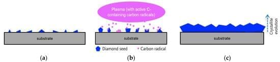
Diamond is a promising material for the biomedical field, mainly due to its set of characteristics such as biocompatibility, strength, and electrical conductivity. Diamond can be synthesised in the laboratory by different methods, is available in the form of plates or films deposited on foreign substrates, and its morphology varies from microcrystalline diamond to ultrananocrystalline diamond. In this review, we summarise some of the most relevant studies regarding the adhesion of cells onto diamond surfaces, the consequent cell growth, and, in some very interesting cases, the differentiation of cells into neurons and oligodendrocytes. We discuss how different morphologies can affect cell adhesion and how surface termination can influence the surface hydrophilicity and consequent attachment of adherent proteins.