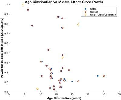
Toggle light / dark theme

Stem cell-gene therapy shows promise in ALS safety trial

Cedars-Sinai investigators have developed an investigational therapy using support cells and a protective protein that can be delivered past the blood-brain barrier. This combined stem cell and gene therapy can potentially protect diseased motor neurons in the spinal cord of patients with amyotrophic lateral sclerosis, a fatal neurological disorder known as ALS or Lou Gehrig’s disease.

In the first trial of its kind, the Cedars-Sinai team showed that delivery of this combined treatment is safe in humans. The findings were reported today in the peer-reviewed journal Nature Medicine.

“Using stem cells is a powerful way to deliver important proteins to the brain or spinal cord that can’t otherwise get through the ,” said senior and corresponding author Clive Svendsen, Ph.D., professor of Biomedical Sciences and Medicine and executive director of the Cedars-Sinai Board of Governors Regenerative Medicine Institute. “We were able to show that the engineered stem cell product can be safely transplanted in the human spinal cord. And after a one-time treatment, these cells can survive and produce an important protein for over three years that is known to protect that die in ALS.”

A Promising Therapy for Hard-To-Treat Depression: Deep Brain Stimulation

A study finds that deep brain stimulation to areas of the brain associated with reward and motivation could be used as a potential treatment for depression.

According to researchers at the University of Texas Health Science Center at Houston, deep brain stimulation (DBS) to the superolateral branch of the medial forebrain bundle (MFB), which is linked to motivation and reward, revealed metabolic brain changes over a 12-month period following DBS implantation. This makes it a potent potential therapy for treatment-resistant depression.

The study’s findings, which included 10 patients, were published in the journal Molecular Psychiatry.

New discovery: Synapse hiding in the mice brain may advance our understanding of neuronal communication

The latest study focused on serotonin receptors in the brain, crucial for memory, fear, and attentiveness.

Scientists have discovered a new type of synapse hiding in the brains of mice. Researchers at HHMI’s Janelia Research Campus.

“This special synapse represents a way to change what is being transcribed or made in the nucleus, and that changes whole programs,” said David Clapham, Janelia’s senior group leader.

“The effects in the cell are not just short-term — some can be long-term,” added the study’s lead author.


Whitehoune/iStock.

Researchers at HHMI’s Janelia Research Campus in Ashburn, Virginia, have found this new kind of synapse in the tiny hairs on the surface of mice neurons, according to a press release published by the institute on Thursday.

When Our Eyes Move During REM Sleep, We’re Gazing at Things in the Dream World

When our eyes move during REM sleep, we’re looking at things in the dream world our brains have created, according to a new study by researchers at the University of California, San Francisco (UCSF). The findings shed light not only on how we dream, but also on how our imaginations work.

REM sleep, which is named for the rapid eye movements associated with it, has been known since the 1950s to be the phase of sleep when dreams occur. But the purpose of the eye movements has remained a matter of much mystery and debate.

REM sleep first occurs about 90 minutes after falling asleep. Your eyes rapidly move from side to side behind closed eyelids. Mixed frequency brain wave activity becomes closer to that seen in wakefulness. Your breathing becomes faster and irregular, and your heart rate and blood pressure increase to near waking levels. Although some can also occur in non-REM sleep, most of your dreaming occurs during REM sleep. Your arm and leg muscles become temporarily paralyzed, which prevents you from acting out your dreams. You sleep less of your time in REM sleep as you age.

How the Brain Processes Sensory Information From Internal Organs

Summary: A new mouse study provides clues as to how the brain processes sensory information from internal organs, revealing feedback from organs activates different clusters of neurons in the brain stem.

Source: Harvard.

Most of us think little of why we feel pleasantly full after eating a big holiday meal, why we start to cough after accidentally inhaling campfire smoke, or why we are hit with sudden nausea after ingesting something toxic. However, such sensations are crucial for survival: they tell us what our bodies need at any given moment so that we can quickly adjust our behavior.

Axolotls can regenerate their brains, revealing secrets of brain evolution and regeneration

The axolotl (Ambystoma mexicanum) is an aquatic salamander renowned for its ability to regenerate its spinal cord, heart and limbs. These amphibians also readily make new neurons throughout their lives. In 1964, researchers observed that adult axolotls could regenerate parts of their brains, even if a large section was completely removed. But one study found that axolotl brain regeneration has a limited ability to rebuild original tissue structure.

So how perfectly can ’s regenerate their brains after injury?

As a researcher studying regeneration at the cellular level, I and my colleagues in the Treutlein Lab at ETH Zurich and the Tanaka Lab at the Institute of Molecular Pathology in Vienna wondered whether axolotls are able to regenerate all the different in their brain, including the connections linking one brain region to another. In our recently published study, we created an atlas of the cells that make up a part of the axolotl brain, shedding light on both the way it regenerates and brain evolution across species.

Axolotls Can Regenerate Their Brains

Summary: Axolotls have the ability to regenerate brain areas following an injury. Researchers have mapped cell types and genes associated with neurodegeneration in the axolotl brain, discovering some similarities in the human brain. The findings could pave the way for new neurodegenerative therapies.

Source: The Conversation.

The axolotl (Ambystoma mexicanum) is an aquatic salamander renowned for its ability to regenerate its spinal cord, heart and limbs. These amphibians also readily make new neurons throughout their lives. In 1964, researchers observed that adult axolotls could regenerate parts of their brains, even if a large section was completely removed. But one study found that axolotl brain regeneration has a limited ability to rebuild original tissue structure.

HBP study explores mechanisms that underlie disorders of consciousness

One of the greatest challenges in the field of neurology and intensive care medicine is correctly diagnosing the level of consciousness of a patient in coma due to severe brain injury. Scientists of the Human Brain Project (HBP) now have explored new techniques that may pave the way to better tell apart two different neurological conditions.

Their findings, published in the journal eLife, reveal important information on the mechanisms of disorders of consciousness.

The team of researchers from University of Liège, GIGA Consciousness Research Unit and Coma Science Group and CHU de Liège (Belgium), Universitat Pompeu Fabra (Spain), Vrije Universiteit Amsterdam (Netherlands), and others, assessed brain functional network states as a marker of consciousness to potentially distinguish patients in the unresponsive wakefulness syndrome (UWS) and minimally conscious state (MCS).