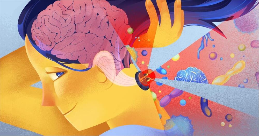
This study explored spatiotemporal progression patterns of striatal dopamine availability and regional brain volume based on cognitive status among patients with Parkinson disease:
Background and Objectives.

Is our brain responsible for how we react to people who are different from us? Why can’t people with autism tell lies? How does the brain produce empathy? Why is imitation a fundamental trait of any social interaction? What are the secret advantages of teamwork? How does the social environment influence the brain? Why is laughter different from any other emotion?
This course is aimed at deepening our understanding of how the brain shapes and is shaped by social behavior, exploring a variety of topics such as the neural mechanisms behind social interactions, social cognition, theory of mind, empathy, imitation, mirror neurons, interacting minds, and the science of laughter.
Serious Science experts from leading universities worldwide answer these and other questions. This course offers a range of scientific perspectives on classical philosophical problems in ethics. It is comprised of 10 lectures filmed from 2014 to 2020. If you have any questions or comments on the content of this course, please write to us at [email protected].
Follow us:
We are on Patreon: / seriousscience.
Facebook — / serious.science.org.
Twitter — / scienceserious.
YouTube — / seriousscience.
Instagram — / serious.science


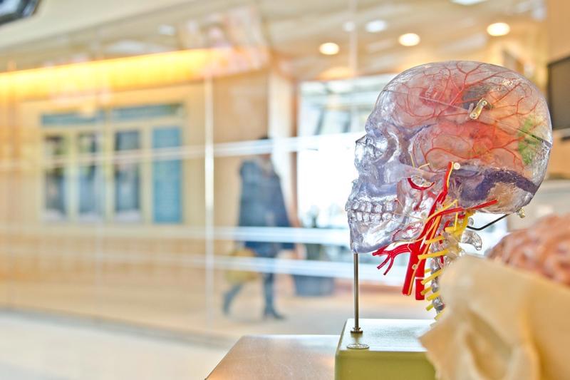
Our willingness to help others is governed by a specific brain region pinpointed by researchers in a study of patients with brain damage to that region.
Learning about where in the brain “helping” decisions are made is important for understanding how people might be motivated to tackle large global challenges, such as climate change, infectious disease and international conflict. It is also essential for finding new approaches to treating disorders of social interactions.
The study, published in Nature Human Behaviour, was carried out by researchers at the University of Birmingham and the University of Oxford, and shows for the first time how a region called the ventromedial prefrontal cortex (vmPFC) has a critical role in helping, or “prosocial” behaviors.

In the future, getting help for depression might involve a quick brain scan to find the most effective treatment for you.
An analysis of brain activity during rest and while undertaking specific tasks among a large group of people with depression and anxiety has identified six distinct types of brain activity patterns, symptoms, and responses to treatment.
The team from the US and Australia who conducted the study also determined treatments that are more likely to work for some of these categories. This means doctors could potentially match patients with the best therapies based on how their brains function.
Scientists have achieved groundbreaking brain mapping using ultrasound, detailed in a May 2024 Science Translational Medicine paper. Led by Richard Andersen and Charles Liu, they developed a non-invasive method to monitor brain activity with unprecedented clarity. This innovation, employing an ‘acoustically transparent’ skull window, allows real-time observation of neuronal and blood flow dynamics. The technique promises new insights into brain function and potential advancements in treating neurological disorders, marking a transformative milestone in neuroscience.

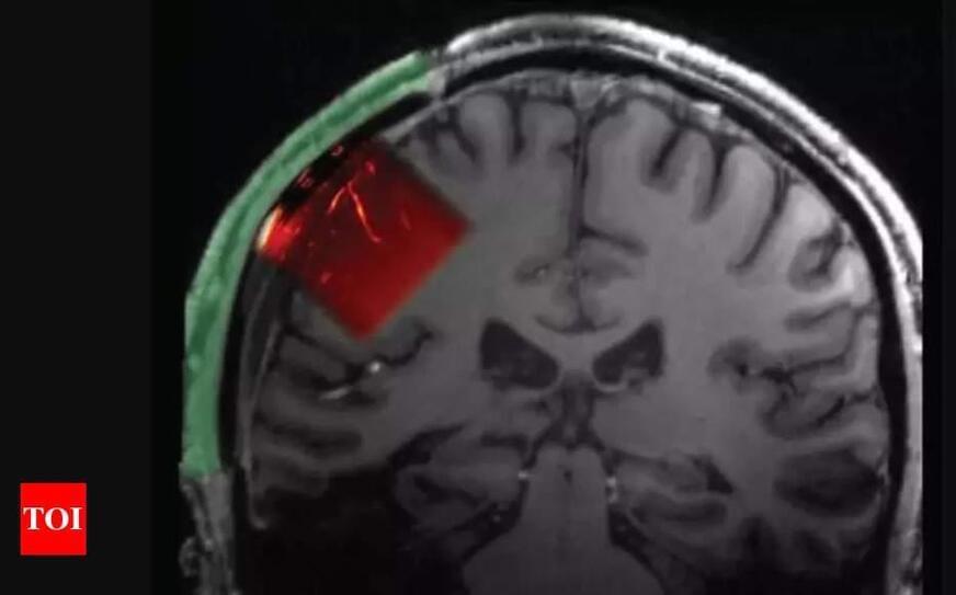
The ability to communicate using only your thoughts might sound like the stuff of science fiction. But for people who don’t have the ability to speak or move due to injury or disease, there’s great hope that this may one day be possible using brain-computer interfaces (BCIs) that can “read” relevant brain signals and translate them into written or spoken words. A research team has made a preliminary advance in this direction by showing for the first time that a computerized brain implant can decode internal speech with minimal training.
In the new NIH-supported study, researchers implanted such a device in a brain area known to be important for representing spoken words called the supramarginal gyrus in two people with tetraplegia, a condition marked by full body paralysis from the neck down due to cervical spinal cord injury. The researchers found that the device could decode several words the participants “spoke” only in their minds. While we are far from using such a device to decode whole sentences or even phrases, and the exact mechanisms of internal speech are still under study, the findings, reported in Nature Human Behavior, are notable because it had been unclear whether the brain signals involved in thinking words could be reproducibly translated.
The findings come from a team led by Richard Andersen at the California Institute of Technology, Pasadena, CA, and Sarah Wandelt, now at the Feinstein Institutes for Medical Research in Manhasset, NY, and the study was supported by the NIH Brain Research Through Advancing Innovative Neurotechnologies® (BRAIN) Initiative Research Opportunities in Humans program. Though earlier research had shown that brain implants could decode vocalized, attempted, and mimed speech, it had yet to be seen whether internal speech could be similarly decoded.
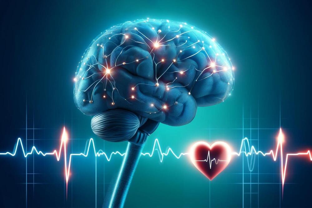
Study suggests heart rate may be a useful tool to determine where to stimulate the brains of individuals with depressive disorders when brain scans aren’t available.
New research suggests a common brain network exists between heart rate deceleration and depression. By evaluating data from 14 people with no depression symptoms, the team of researchers at Brigham and Women’s Hospital, a founding member of the Mass General Brigham healthcare system, found stimulating some parts of the brain linked to depression with transcranial magnetic stimulation (TMS), also affected heart rate, suggesting clinicians may be able to target those areas without the use of brain scans that aren’t widely available. The findings were published recently in the journal Nature Mental Health.
Heart-Brain Coupling and TMS.