The provided text outlines Joscha Bach theories regarding the nature of synthetic consciousness and the limitations of modern science. Bach posits that human experience is not a direct interaction with reality, but rather a simulated world model constructed by the brain internal software. He defines intelligence as the capacity to build these models in novel environments, suggesting that current artificial intelligence remains incomplete because it lacks genuine self-understanding. Furthermore, he challenges the narrow focus of contemporary academia and traditional neuroscience, arguing that minds are complex information-processing systems that cannot be explained by neural connections alone. Ultimately, these sources present a computational framework for understanding the self as a functional narrative rather than a mystical or purely physical entity.
Category: neuroscience – Page 48
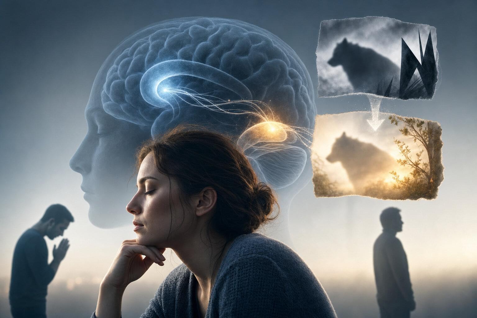
Your brain doesn’t forget when you forgive—it does something far more surprising with those painful memories
Forgiving someone might not erase painful memories, but it can subtly update them, making past hurts feel less upsetting. It’s less “forgive and forget,” and more “forgive and update.”
Psychologists have long known that forgiveness is crucial for healing rifts and keeping social bonds strong. Folk wisdom even advises us to “forgive and forget” after a wrong, implying that saying you forgive someone should make the bad memory vanish.
But forgiving doesn’t actually make you forget, notes Duke neuroscientist Felipe de Brigard: “When you forgive someone for a wrongdoing, you don’t forget the event. But once you forgive, the memory doesn’t hurt as much.” Indeed, past studies hinted that forgiving someone can blunt the memory of their misdeed. What hasn’t been clear is how that happens in the brain. Is the memory simply erased, or does it get rewritten?

Childhood trauma predicts higher risk of combined mental and physical illness in later life
Researchers modeled the specific dosage of trauma to highlight an escalating relationship between the sheer volume of trauma and later health vulnerabilities. Small amounts of childhood adversity corresponded to relatively modest increases in health risks. However, once a person’s trauma score passed four distinct adverse experiences, the upward trajectory of their health risk accelerated rapidly.
The researchers also investigated the stepping stones connecting early trauma to later disease onset. Using a statistical technique called mediation analysis, they looked for intermediate health issues that acted as bridges over the span of a lifetime. They found that developing either a single physical illness or isolated depression in early adulthood often served as an indirect pathway to combined disease in older age.
For individuals with the highest amounts of early trauma, early-onset depression played a particularly strong bridging role. An initial diagnosis of depression frequently paved the way for additional physical conditions as time went on. These findings align with biological theories suggesting that severe childhood stress permanently disrupts the body’s immune regulation and stress hormone pathways.
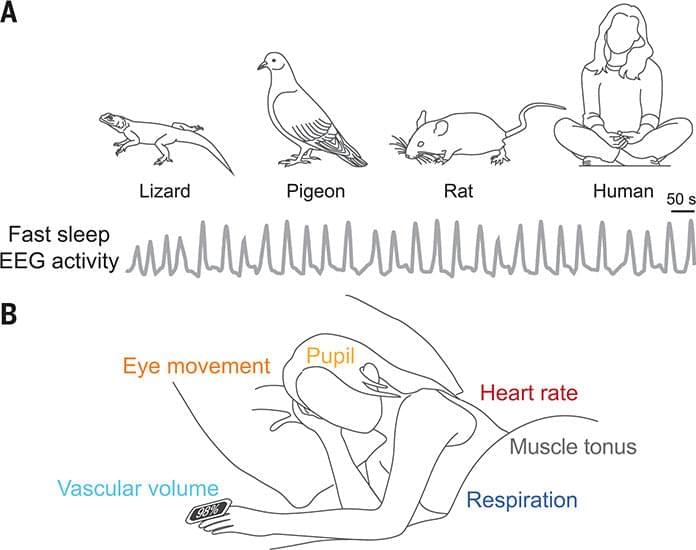
The oscillatory biology of sleep: Linkage to dementia
During wakefulness, neuromodulators operate largely independently to support behavior and cognition. By contrast, sleep reorganizes their activity into a coordinated brain rhythm. During sleep, the major neuromodulators—norepinephrine, acetylcholine, serotonin, and dopamine—exhibit synchronized fluctuations with a periodicity of ~50 seconds. These oscillations appear as recurrent bursts of fast (10 to 30 hertz) electroencephalography activity and are phase-coupled to cerebrospinal fluid flow. Neuromodulators are vasoactive agents and drive slow vasomotion, which provide the mechanical force that supports glymphatic clearance of metabolic waste. Disruption of neuromodulator signaling, as seen in psychiatric disorders, cardiovascular disease, aging, or with commonly prescribed drugs, impairs clearance of neurotoxic proteins, including amyloid-β and tau.
How the Brain Remembers Trauma Differently: Understanding Traumatic Memory
Traumatic memories are a complicated psychological phenomenon, where some experiences are never forgotten but can only be remembered in fragments. Traumatic memories are not as complete or coherent as regular memories. Even if its details are absent, the actual event can still make a strong impression. Trauma is the result of an extremely stressful, frightening or upsetting event that is hard to cope with or feel we have no control over. These experiences may be a one-time thing or repeated over time.
Optimize Brain Health And Longevity: Tommy Wood, MD, PhD
Join us on Patreon! / michaellustgartenphd.
Discount Links/Affiliates:
Blood testing (where I get the majority of my labs, for those who blood test with Quest): https://www.ultalabtests.com/partners… those who blood test with LabCorp: https://www.anrdoezrs.net/click-10161… At-Home Metabolomics: https://www.iollo.com?ref=michael-lus… Use Code: CONQUERAGING At Checkout Clearly Filtered Water Filter: https://get.aspr.app/SHoPY Epigenetic, Telomere Testing: https://trudiagnostic.com/?irclickid=… Use Code: CONQUERAGING NAD+ Quantification: https://www.jinfiniti.com/intracellul… Use Code: ConquerAging At Checkout Oral Microbiome: https://www.bristlehealth.com/?ref=mi… Enter Code: ConquerAging SiphoxHealth Blood Testing (ApoB, GrimAge): https://siphoxhealth.com/mlustgarten Green Tea: https://www.ochaandco.com/?ref=fqbtflod Use Code: ML10OFF Diet Tracking: https://shareasale.com/r.cfm?b=139013… If you’d like to support the channel, you can do that with the website, Buy Me A Coffee: https://www.buymeacoffee.com/mlhnrca Conquer Aging Or Die Trying Merch! https://my-store-d4e7df.creator-sprin…
Blood Testing Essentials (Biological Age, CVD-Risk, Kidney Health and Function):
PhenoAge (Biological Age): https://www.ultalabtests.com/partners…
Risk-weighted ApoB (a better CVD predictor than LDL, non-HDL cholesterol, and ApoB): https://www.ultalabtests.com/partners…
Kidney health and function: https://www.ultalabtests.com/partners…