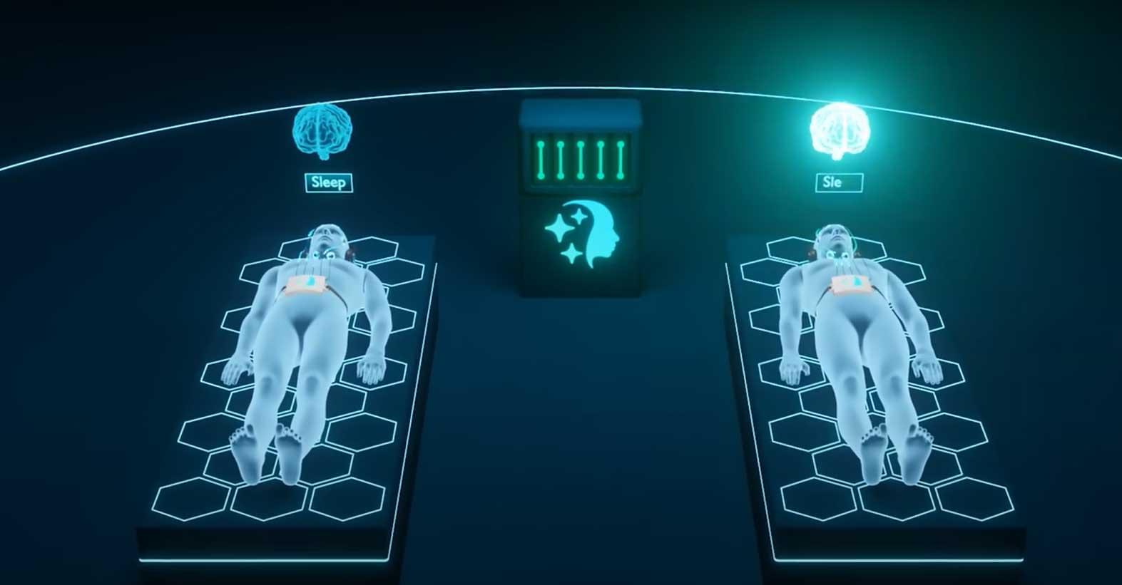
Participants successfully exchanged information through lucid dreams.
Scientists discovered that a species of fungus can sense its surroundings and make strategic decisions.
A new study suggests that the fungus Phanerochaete velutina might have a surprising ability — recognizing shapes and adjusting its growth strategy accordingly.
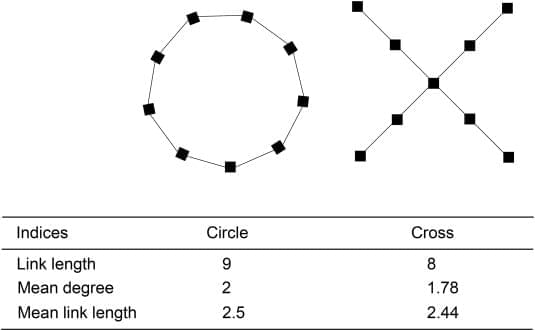
Researchers from Tohoku University conducted experiments where the fungus was placed in different spatial arrangements and observed how it spread. Rather than expanding indiscriminately, the mycelium formed connections, retracted excess strands, and focused its foraging in strategic directions.
In circular arrangements, tendrils avoided the center, while in cross-shaped formations, the outermost blocks served as primary hubs for exploration.
This behavior hints at a level of perception and decision-making previously unrecognized in fungi.
The findings add to growing evidence that fungi exhibit a form of primitive intelligence, capable of memory, learning, and problem-solving. Scientists believe this research could expand our understanding of cognition in non-animal organisms, as well as inspire innovations in bio-computing.
The ability of fungal networks to process information and optimize resource allocation without a brain challenges conventional ideas about intelligence. As studies continue, fungi may offer fascinating insights into decentralized decision-making and ecological adaptation.

Mashour is one of a small set of clinicians and scientists trying to change that. They are increasingly bringing the tools of neuroscience into the operating room to track the brain activity of patients, and testing out anesthesia on healthy study participants. These pioneers aim to learn how to more safely anesthetize their patients, tailoring the dose to individual patients and adjusting during surgery. They also want to better understand what governs the transitions between states of consciousness and even hope to crack the code of coma.
Your brain on anesthesia
Today’s anesthetic arsenal eschews Morton’s original formula for newer, safer drugs. These include ether-based inhalants such as sevoflurane and isoflurane, and the widely used, intravenous anesthetic propofol, all of which wear off faster than early ether-based anesthetics, enabling quicker recovery. (They are also less likely to cause fires and explosions in the operating room, a regular occurrence through the first half of the 20th century.) Despite these improvements, the risks associated with excessive sedation remain high. Depending on the complexity and length of surgery, between 17 and 43 percent of patients may have cognitive problems, typically in memory and executive functions.1 These typically last only one to two weeks after surgery, but few rigorous studies have examined changes in cognitive function in the general population beyond six months after surgery.

Summary: A new study suggests that ChatGPT’s responses in psychotherapy scenarios are often rated higher than those written by human therapists. Researchers found that participants struggled to distinguish between AI-generated and therapist-written responses in couple’s therapy vignettes. ChatGPT’s responses were generally longer and contained more nouns and adjectives, providing greater contextualization.
This additional detail may have contributed to higher ratings on core psychotherapy principles. The findings highlight AI’s potential role in therapeutic interventions while raising ethical and practical concerns about its integration into mental health care. Researchers emphasize the need for professionals to engage with AI developments to ensure responsible oversight.

Given that disparate mind/body views have interfered with interdisciplinary research in psychoanalysis and neuroscience, the mind/body problem itself is explored here. Adding a philosophy of mind framework, problems for both dualists and physicalists are presented, along with essential concepts including: independent mental causation, emergence, and multiple realization. To address some of these issues in a new light, this article advances an original mind/body account—Diachronic Conjunctive Token Physicalism (DiCoToP). Next, puzzles DiCoTop reveals, psychoanalytic problems it solves, and some empirical evidence accrued for views consistent with DiCoToP are presented. In closing, this piece challenges/appeals for neuroscience research to gain evidence for (or against) the DiCoToP view.


Most neuroscience research carried out up to date has primarily focused on neurons, the most renowned type of cell in the human brain. As a result, the unique functions of other brain cell types are less understood and have often been entirely overlooked.
Researchers at Instituto Cajal (CSIC), the Autonomous University of Madrid and Institute de Salud Carlos III recently carried out a study aimed at better understanding the contributions of astrocytes, a class of star-shaped glial cells found in the brain and spinal cord, to key mental functions. Their findings, published in Nature Neuroscience, unveiled the existence of astrocytic ensembles, specialized astrocyte subsets that appear to be active during reward-driven behaviors.
“It is known that astrocytes are a heterogeneous cell type in their molecular and gene expression signatures, morphology and origin,” Marta Navarrete, senior author of the paper, told Medical Xpress.

Our brain and eyes can play tricks on us—not least when it comes to the expanding hole illusion. A new computational model developed by Flinders University experts helps to explain how cells in the human retina make us “see” the dark central region of a black hole graphic expand outwards.
In a new article posted to the arXiv preprint server, the Flinders University experts highlight the role of the eye’s retinal ganglion cells in processing contrast and motion perception—and how messages from the cerebral cortex then give the beholder an impression of a moving or “expanding hole.”
“Visual illusions provide valuable insights into the mechanisms of human vision, revealing how the brain interprets complex stimuli,” says Dr. Nasim Nematzadeh, from the College of Science and Engineering at Flinders University.
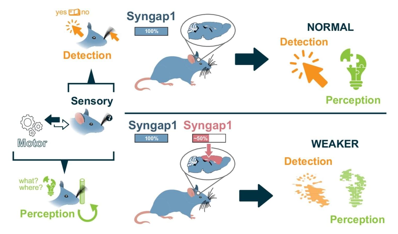
The SYNGAP1 gene, which supports the production of a protein called SynGAP (Synaptic Ras GTPase-Activating Protein), is known to play a key role in supporting the development of synapses and neural circuits (i.e., connections between neurons). Mutations in this gene have been linked to various learning disabilities, including intellectual disabilities, speech and language delays, autism spectrum disorder (ASD), and epilepsy.
Researchers at the Herbert Wertheim UF Scripps Institute for Biomedical Innovation & Technology recently carried out a study aimed at better understanding the genetic mechanisms via which the SYNGAP1 gene contributes to healthy cognitive function. Their findings, published in Nature Communications, suggest that the autonomous expression of this gene in the cortical excitatory neurons of mice promotes the animals’ cognitive abilities via the assembly of long-range neural circuits integrating sensory and motor information.
“Our paper builds on our ongoing research into how major risk genes for mental health disorders, including autism, regulate brain organization and function,” Gavin Rumbaugh, senior author of the paper, told Medical Xpress. “The field knows the major risk genes that directly contribute to cognitive and behavioral impairments that lead to diagnosable forms of autism and related neuropsychiatric disorders in humans.
In cognitive warfare truth becomes lies good becomes evil.
Provided to YouTube by Routenoteall the collapsing stars · sarochi · sarochi (ken)sarochi℗ SAROCHIReleased on: 2025–02-05Auto-generated by YouTube.