“We know that as little as 10 minutes of walking can improve your mood, getting that bubble bath with the dopamine, serotonin, endorphins going. Anybody can do that.”


A new neuroimaging study from China has found that an eight-week course of bright light therapy helped reduce depressive symptoms in individuals with subthreshold depression. The treatment also altered dynamic functional connectivity in several brain regions associated with mood regulation. The study was published in the Journal of Affective Disorders.
Subthreshold depression refers to the presence of depressive symptoms that are clinically relevant but do not meet the full diagnostic criteria for major depressive disorder. Individuals with subthreshold depression may experience persistent sadness, fatigue, sleep disturbances, or concentration problems, but with fewer symptoms or a shorter duration than required for a formal diagnosis.
Despite being “subthreshold,” the condition can impair daily functioning and reduce quality of life. It is also linked to an increased risk of developing major depression in the future. Subthreshold depression is common—especially among adolescents, older adults, and individuals with chronic illnesses—and it often goes undiagnosed and untreated because the symptoms are perceived as mild or situational. However, research shows that even mild depressive symptoms can negatively affect social relationships, job performance, and physical health.
As researchers work to improve treatment of Alzheimer’s disease, new research by UCLA Health identified a candidate drug that reduces levels of a toxic form of a protein in the brain caused by the disease and improved memory in mice by boosting production of a protective protein.
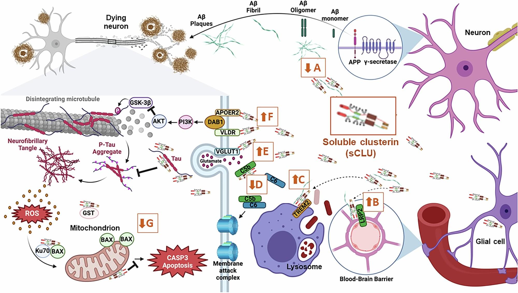
In a study published in npj Drug Discovery, UCLA Health researchers targeted the protein clusterin (CLU), which is crucial in preventing the build-up of amyloid-beta plaques and tau proteins that disrupt communication between brain cells and lead to memory impairment—a hallmark symptom of Alzheimer’s disease.
More than a decade ago, a variant of the gene that encodes clusterin was identified as the third strongest genetic risk factor for late-onset Alzheimer’s disease. It was recently reported that increased CLU protein could provide protection against Alzheimer’s disease and cognitive decline.

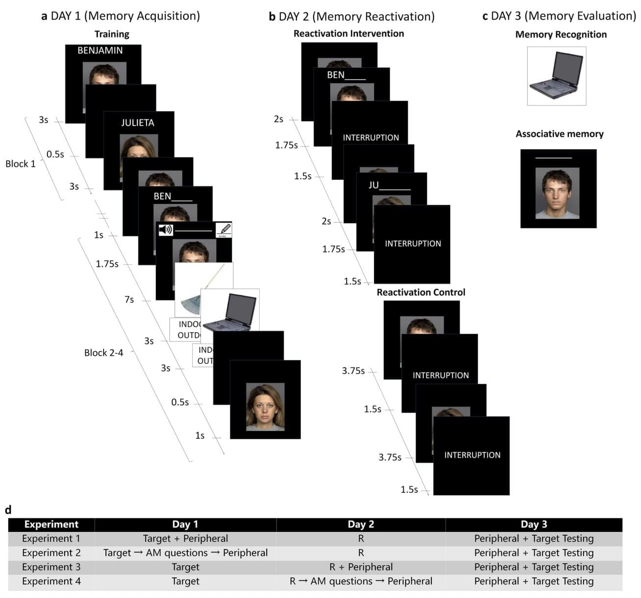
Most humans can recall specific events and past experiences for long periods of time. This capability, referred to as episodic memory, is known to be in great part supported by the activity of neurons in the hippocampus and medial temporal lobe.
Past neuroscience and psychology studies consistently found that episodic memory is associative. This essentially means that remembering one past event, for instance a graduation, can in many cases prompt people to also remember other related events, such as a party that celebrated the graduation.
Researchers at Biología Molecular y Neurociencias (IFIByNE)-CONICET and the University of Buenos Aires recently carried out a new study exploring the possibility that the reactivation of specific episodic memories does not only help to strengthen those memories, but also the memories of other related events or experiences.
New studies stemming from the Armamentarium consortium outline findings that advance tools based on Adeno-associated virus (AAV) vectors. An announcement about the work explains how an AAV “acts like a shuttle capable of transporting specially designed DNA into the cell.”
Two of the studies on these AAV tools were conducted by collaborative teams organized by Xiangmin Xu, Ph.D., UC Irvine Chancellor’s Professor of anatomy and neurobiology and director of the campus’s Center for Neural Circuit Mapping.
“This Armamentarium’s collection of work enables new tools that help to deepen our understanding of the human central nervous system structure and function,” says Xu. “Our own brain-targeting technology could help treat Alzheimer’s disease and many other neurological disorders.”