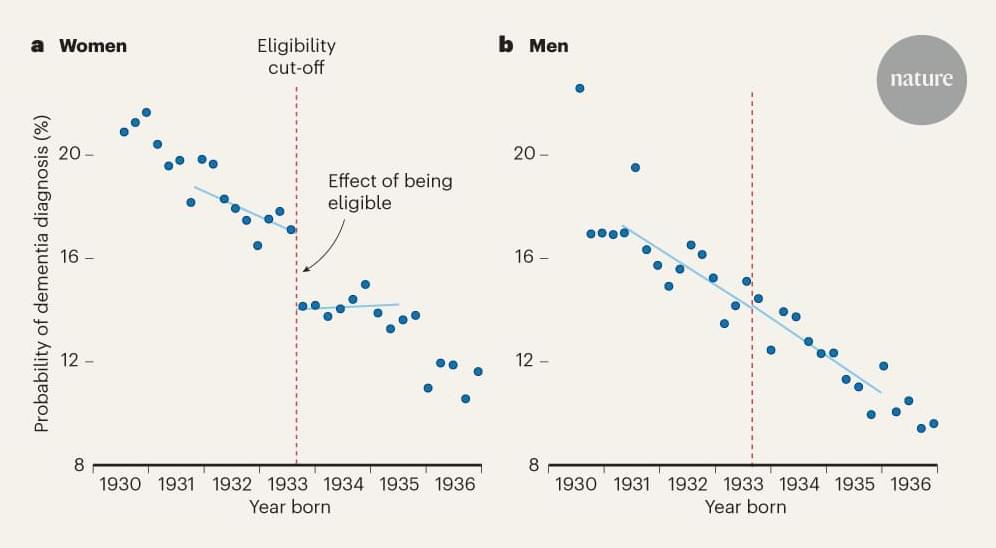
Large-scale analyses of electronic health data suggest that the herpes zoster vaccine could protect against dementia — but it’s not yet clear how.


A new study reveals that how your brain reacts to food purchasing decisions can be used to determine your political affiliation with almost 80% accuracy.
Researchers from Iowa State University, the University of Kansas Medical Center, Oklahoma State University, and the University of Exeter in England used brain imaging techniques to examine adults purchasing eggs and milk with various prices and produced in different ways.
Interestingly, the purchases did not significantly differ by the adults’ political party. What differed by political affiliation, were the areas of the brain that were active during the purchases. The study is published in the journal Politics and the Life Sciences.
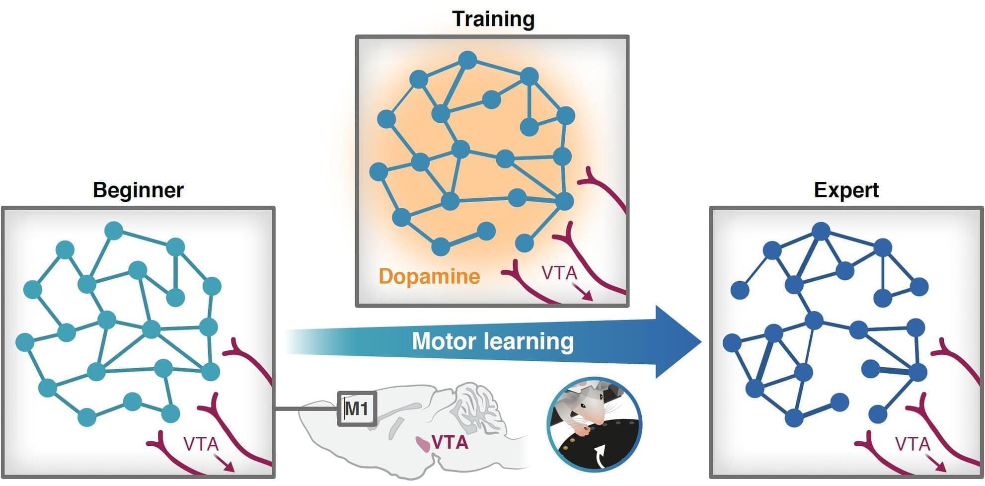
A new interdisciplinary study by researchers from the Ruth and Bruce Rappaport Faculty of Medicine and the Andrew and Erna Viterbi Faculty of Electrical and Computer Engineering at the Technion reveals a surprising insight: local release of dopamine—a molecule best known for its role in the brain’s reward system—is a key factor in acquiring new motor skills

Scientists studying mice have uncovered a delicate protein rivalry in the brain that, when thrown off balance, may cause autism-like behaviors.
This discovery opens up a potentially powerful new path for autism treatment by targeting how nerve signals are regulated at the molecular level.
Protein imbalance tied to autism symptoms.
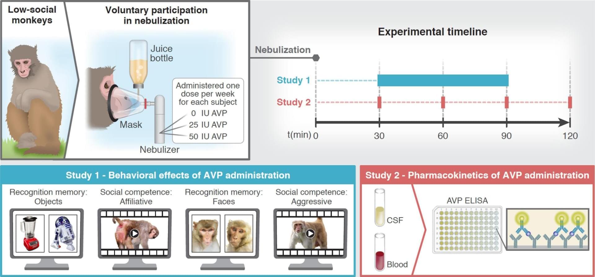
I very much enjoyed reading this nicely done preclinical study on using nebulized vasopressin to improve social cognition in low-sociality rhesus monkeys. Reading about their study design in particular was highly informative/educational! #preclinical #medicine #biomedicine
Low cerebrospinal (CSF) arginine vasopressin (AVP) concentration is a biomarker of social impairment in low-social monkeys and children with autism, suggesting that AVP administration may improve primate social functioning. However, AVP administration also increases aggression, at least in “neurotypical” animals with intact AVP signaling. Here, we tested the effects of a voluntary drug administration method in low-social male rhesus monkeys with high autistic-like trait burden. Monkeys received nebulized AVP or placebo, using a within-subjects design. Study 1 (N = 8) investigated the effects of AVP administration on social cognition in two tests comparing responses to social versus nonsocial stimuli. Test 1: Placebo-administered monkeys lacked face recognition memory, whereas face recognition memory was “rescued” following AVP administration.
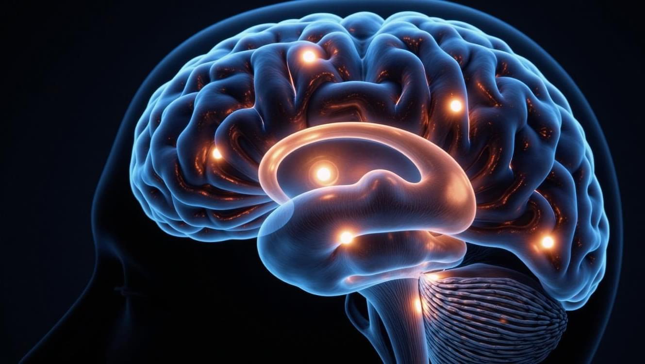
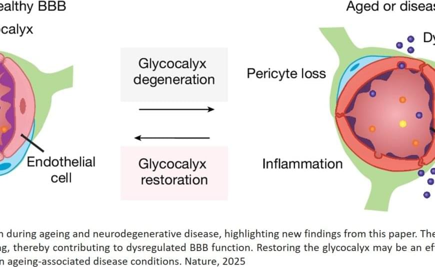
In a study in aging mice, the first author has uncovered striking age-related changes in the sugary coating – called the glycocalyx – on cells that form the blood-brain barrier, a structure that protects the brain by filtering out harmful substances while allowing in essential nutrients.
“The glycocalyx is like a forest,” the author explains. “In young, healthy brains, this forest is lush and thriving. But in older brains, it becomes sparse, patchy, and degraded.”
These age-related changes to the glycocalyx weaken the blood-brain barrier, the author found. As the barrier becomes leaky with age, harmful molecules can infiltrate the brain, potentially fueling inflammation, cognitive decline, and neurodegenerative diseases.
The results were striking: In older mice, bottlebrush-shaped, sugar-coated proteins called mucins, a key component of the glycocalyx, were significantly reduced. This thinning of the glycocalyx correlated with increased permeability of the blood-brain barrier and heightened neuroinflammation.
When the team reintroduced those critical mucins in aged mice, restoring a more “youthful” glycocalyx, they improved the integrity of the blood-brain barrier, reduced neuroinflammation, and measurably improved cognitive function.
“Modulating glycans has a major effect on the brain – both negatively in aging, when these sugars are lost, and positively, when they are restored,” the lead says. “This opens an entirely new avenue for treating brain aging and related diseases.”
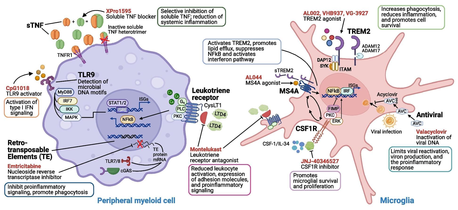
Immune mechanisms play a fundamental role in Alzheimer’s disease (AD) pathogenesis, suggesting that approaches which target immune cells and immunologically relevant molecules can offer therapeutic opportunities beyond the recently approved amyloid beta monoclonal therapies. In this review, we provide an overview of immunomodulatory therapeutics in development, including their preclinical evidence and clinical trial results. Along with detailing immune processes involved in AD pathogenesis and highlighting how these mechanisms can be therapeutically targeted to modify disease progression, we summarize knowledge gained from previous trials of immune-based interventions, and provide a series of recommendations for the development of future immunomodulatory therapeutics to treat AD.