Toggle light / dark theme

The same technique could also be applied to studies of brain damage, Ruetz said. “Neural stem cells in the subventricular zone are also in the business of repairing brain tissue damage from stroke or traumatic brain injury.”

The glucose transporter connection “is a hopeful finding,” Brunet said. For one, it suggests not only the possibility of designing pharmaceutical or genetic therapies to turn on new neuron growth in old or injured brains, but also the possibility of developing simpler behavioral interventions, such as a low carbohydrate diet that might adjust the amount of glucose taken up by old neural stem cells.

The researchers found other provocative pathways worthy of follow-up studies. Genes relating to primary cilia, parts of some brain cells that play a critical role in sensing and processing signals such as growth factors and neurotransmitters, also are associated with neural stem cell activation. This finding reassured the team that their methodology was effective, partly because unrelated previous work had already discovered associations between cilia organization and neural stem cell function. It is also exciting because the association with the new leads about glucose transmission could point toward alternative avenues of treatment that might engage both pathways, Brunet said.

Send us a text.

Put simply, the brain is not too warm or wet for consciousness to exist as a wave that connects with the universe.

For decades, Penrose has been working with anesthesiologist Stuart Hameroff on a theory of consciousness called Orchestrated Objective Reduction (Orch OR). Penrose primarily handles the physics of Orch OR, whereas Hameroff handles the biology. Their theory addressed serious gaps in established scientific frameworks spanning physics, neuroscience and psychology. All, some or none of the hypotheses in this theory might prove out experimentally. See the paper below as a step towards proof.

Researchers from the Color and Food Quality group at the Faculty of Pharmacy, University of Seville, in partnership with Dr. Marina Ezcurra’s team at the University of Kent (UK), have demonstrated that the carotenoid phytoene extends the lifespan of the nematode Caenorhabditis elegans. Additionally, it delays the onset of paralysis linked to amyloid plaque formation in an Alzheimer’s disease model.

Specifically, increases in longevity of between 10 and 18.6% and decreases in the proteotoxic effect of plaques of between 30 and 40% were observed. The studies, which form part of Ángeles Morón Ortiz’s doctoral thesis, tested pure phytoene and extracts rich in this carotenoid obtained from microalgae.

According to Dr. Paula Mapelli Brahm, “These are very exciting preliminary results, so we are looking for funding to continue this line of research and to find out by what mechanisms these effects are produced.”

A new brain-mapping tool just dropped!


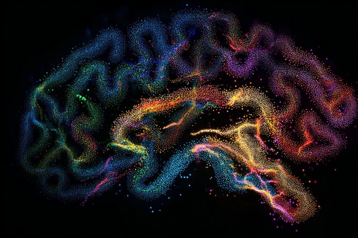
LA JOLLA—Scientists at the Salk Institute are unveiling a new brain-mapping neurotechnology called Single Transcriptome Assisted Rabies Tracing (START). The cutting-edge tool combines two advanced technologies—monosynaptic rabies virus tracing and single-cell transcriptomics—to map the brain’s intricate neuronal connections with unparalleled precision.

Using the technique, the researchers became the first to identify the patterns of connectivity made by transcriptomic subtypes of inhibitory neurons in the cerebral cortex. They say having this ability to map the connectivity of neuronal subtypes will drive the development of novel therapeutics that can target certain neurons and circuits with greater specificity. Such treatments could be more effective and produce fewer side effects than current pharmacological approaches.

The study, published on September 30, 2024, in Neuron, is the first to resolve cortical connectivity at the resolution of transcriptomic cell types.

Source: Allen Institute.

The BRAIN Initiative® Cell Atlas Network (BICAN) has launched its first major data release, marking a significant milestone in the ambitious effort to map the whole human brain.

The data, accessible through the BICAN Rapid Release Inventory, includes single-cell and single-nucleus transcriptomic and epigenomic profiles from humans, mice, and 10 other mammalian species.

New steps have been taken towards a better understanding of the immediate and long-term impact of COVID-19 on the brain in the UK’s largest study to date.

Published in Nature Medicine, the study from researchers led by the University of Liverpool alongside King’s College London and the University of Cambridge as part of the COVID-CNS Consortium shows that 12–18 months after hospitalisation due to COVID-19, patients have worse cognitive function than matched control participants. Importantly, these findings correlate with reduced brain volume in key areas on MRI scans as well as evidence of abnormally high levels of brain injury proteins in the blood.

Strikingly, the post-COVID cognitive deficits seen in this study were equivalent to twenty years of normal ageing. It is important to emphasise that these were patients who had experienced COVID, requiring hospitalisation, and these results shouldn’t be too widely generalised to all people with lived experience of COVID. However, the scale of deficit in all the cognitive skills tested, and the links to brain injury in the brain scans and blood tests, provide the clearest evidence to date that COVID can have significant impacts on brain and mind health long after recovery from respiratory problems.

A powerful new analytical tool offers a closer look at how tumor cells “shape-shift” to become more aggressive and untreatable, as shown in a study from researchers at Weill Cornell Medicine and the New York Genome Center.

A tumor cell shape-shifts by changing its cell type or state, thus altering its basic pattern of activity and perhaps even its appearance. This changeability or “plasticity” is a characteristic of cancer that leads to diverse tumor-cell populations and ultimately the emergence of cell types enabling treatment resistance and metastatic spread.

The new tool, described Sept. 24 in a paper in Nature Genetics, can be used to quantify this plasticity in samples of tumor cells. The researchers demonstrated it with analyses of tumor samples from animal models and human patients, identifying, for example, a key transitional cell state in glioblastoma, the most common form of brain cancer.

The FDA has approved a new targeted drug specifically for brain tumors called low-grade gliomas. The drug, vorasidenib, was shown in clinical trials to delay progression of low-grade gliomas that had mutations in the IDH1 or IDH2 genes.

“Although there have been other targeted therapies for the treatment of brain tumors with the IDH mutation, [this one] has been one of the most successful in survival prolongation of brain tumor patients,” said Darell Bigner, MD, PhD, the E. L. and Lucille F. Jones Cancer Distinguished Research Professor and founding director of the Preston Robert Tisch Brain Tumor Center at Duke.

In clinical trials, progression-free survival was estimated to be 27.7 months for people in the vorasidenib group versus 11.1 months for those in the placebo group.