A recent large population study claims to conclusively prove that male and female brains are different. Of course, it does no such thing. As such, Dr Dean Burnett is quite annoyed.
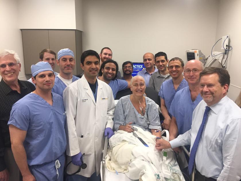
MORGANTOWN — World-leading brain experts at West Virginia University’s Rockefeller Neuroscience Institute are celebrating the historic breakthrough Alzheimer patients around the global have been waiting for.
“For Alzheimer’s, there’s not that many treatments available despite hundreds of clinical trials over the past two decades and billions of dollars spent,” said Dr. Ali Rezai, a neurosurgeon at WVU who led the team of investigators that successfully performed a phase II trial using focused ultrasound to treat a patient with early stage Alzheimer’s.
The WVU team tested the innovative treatment in collaboration with INSIGHTEC, an Israeli medical technology company. Earlier this year, INSIGHTEC was approved by the U.S. Food and Drug Administration to begin a phase II clinical trial of the procedure and selected the WVU Rockefeller Neuroscience Institute as the first site in the United States for the trial.
Samsung TVs are already some of the most popular options for high-end home theater systems, and the company is now using its television-making prowess to help people with disabilities live more normal lives. A new project by a Samsung team in Switzerland could yield the first smart TV that can be controlled with thoughts.
As CNET reports, Samsung has partnered with Swiss scientists to bring the system to life. Called ‘Project Pontis,’ the ultimate goal is to build a brain/software interface that will allow individuals with movement disabilities to control television features like channel switching and volume control with their brains rather than their bodies.

Motivation is such an intangible aspect of the human spirit that we often forget it has very real, neurochemical origins. We admire it in others and strive for it in ourselves (see: every Nike ad ever made), and now we are getting closer to potentially inducing that motivational feeling with drugs.
John Salamone, Ph.D., a professor at the University of Connecticut with a background in neural and behavioral pharmacology, has been working with the drug company Chronos Therapeutics to develop a drug that can restore motivation in people who have lost it — whether that’s due to the symptoms of depression, struggle with disease, or otherwise. He unveiled his early results on rats this week in a presentation at the Society For Neuroscience’s conference in San Diego, where he tells Inverse his board was bustling with activity:
“Basically we stood there for four hours and were busy the entire time,” says Salamone. The reception was overwhelmingly positive, he adds. “We didn’t have anyone say ‘This is crazy! This will never work!’”.
I think aspects of our Universe are conscious. Dark matter or the æther perhaps. Stagnancy is death so change inherently means something is making decisions.
A new scientific concept has recently come to light, which scientists are calling “panpsychism.” Panpsychism says that the universe could be capable of consciousness, which could change everything.
For quite some time, scientists have been working to understand the universe, where it came from, and why we are here. However, they have often come up short until now. The scientist responsible for such a notion is Gregory Matloff, and his ideas are shocking, to say the least.
According to Matloff, a physicist at New York City College of Technology, in his recently published paper, humans could be like the rest of the universe, in substance and in spirit. Futurism reported that a “proto-consciousness field” could extend throughout all space. Basically, in lamens terms, the entire cosmos could be self-aware.
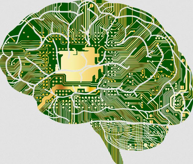
SpiNNaker was built under the leadership of Professor Steve Furber at The University of Manchester, a principal designer of two products that earned the Queen’s Award for Technology —the ARM 32-bit RISC microprocessor, and the BBC Microcomputer.
“The ultimate objective for the project has always been a million cores in a single computer for real time brain modelling applications, and we have now achieved it, which is fantastic.” — Professor Steve Furber, The University of Manchester
Inspired by the human brain, the SpiNNaker is capable of sending billions of small amounts of information simultaneously. The SpiNNaker has a staggering 1 million processors that are able to perform over 200 million actions per second.
Grunya Sukhareva characterized autism nearly two decades before Austrian doctors Leo Kanner and Hans Asperger.

Prolonged social isolation can do severe, long-lasting damage to the brain.