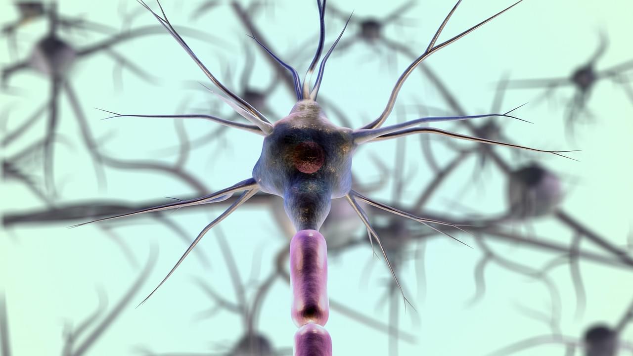
A new computer simulation of how our brains develop and grow neurons has been built by scientists from the University of Surrey. Along with improving our understanding of how the brain works, researchers hope that the models will contribute to neurodegenerative disease research and, someday, stem cell research that helps regenerate brain tissue.
Category: neuroscience – Page 116
What Memories Are Made Of
Posted in neuroscience
The Top 4 Most viewed videos in the FENS Video Contest will be announced on June 10. View and Share to Vote for your favourite!
Description of the video:
This video is about how memories are stored in the brain. We are exploring how with an experiment, called long-term potentiation (LTP), we can modulate the strength of the connection between neurons. Combining filming in the laboratory with the use of bioinformatic tools and animation, we try to illustrate the cellular and molecular basis of this phenomenon.
Author: Tanja Fuchsberger, Department of Physiology, Development and Neuroscience. University of Cambridge. UK
Music: Composed by Juan Manuel Pastor Labrandero (from the band Johnny B. Zero https://www.johnny-b-zero.com/)
Weight-maintaining hormone key to brain-signal transmission. A study more closely links obesity to dementia, finding that leptin, a hormone that helps maintain normal body weight, is associated with better signal-transmitting brain white matter in middle-aged adults.
New research is more closely linking obesity to dementia.
Higher levels of leptin, a hormone that helps maintain normal body weight, is associated with better signal-transmitting brain white matter in middle-aged adults, according to a study by The University of Texas Health Science Center at San Antonio (UT Health San Antonio).
This study explores how the brain processes predictions and what happens to these processes during unconsciousness induced by the anesthetic propofol.
Our brains are constantly making predictions about our surroundings, enabling us to focus on and respond to unexpected events. A recent study explores how this predictive process functions during consciousness and how it changes under general anesthesia. The findings support the idea that conscious thought relies on synchronized communication between basic sensory areas and higher-order cognitive regions of the brain, facilitated by brain rhythms in specific frequency bands.
Previously, members of the research team at The Picower Institute for Learning and Memory at MIT and at Vanderbilt University had described how brain rhythms enable the brain to remain prepared to attend to surprises. Cognition-oriented brain regions (generally at the front of the brain), use relatively low-frequency alpha and beta rhythms to suppress processing by sensory regions (generally toward the back of the brain) of stimuli that have become familiar and mundane in the environment (e.g. your co-worker’s music). When sensory regions detect a surprise (e.g. the office fire alarm), they use faster frequency gamma rhythms to tell the higher regions about it and the higher regions process that at gamma frequencies to decide what to do (e.g. exit the building).
Anesthesia’s Impact on Brain Communication
The new results published Oct. 7 in the Proceedings of the National Academy of Sciences, show that when animals were under propofol-induced general anesthesia, a sensory region retained the capacity to detect simple surprises but communication with a higher cognitive region toward the front of the brain was lost, making that region unable to engage in its “top-down” regulation of the activity of the sensory region and keeping it oblivious to simple and more complex surprises alike.
Pigs are considered useful biomedical models for humans so the implications of such studies sent waves through the field of resuscitation — and bioethics.
Summary: Researchers have developed the first 3D map of gene regulation in the human brain, offering insights into how early brain development influences lifelong mental health. This map, focusing on regions tied to memory and emotional regulation, reveals how chromatin structure controls gene activity, especially during key developmental stages.
These findings may help identify when and where genetic variants linked to autism and schizophrenia disrupt normal development. By understanding these early influences, scientists hope to improve neurodevelopmental disorder research and stem-cell models, potentially paving the way for earlier intervention strategies.
REDWOOD CITY, Calif.—(BUSINESS WIRE)—Researchers at REMspace, a California-based startup, have achieved a historic milestone, demonstrating that lucid dreams could unlock new dimensions of communication and humanity’s potential. Using specially designed equipment, two individuals successfully induced lucid dreams and exchanged a simple message.
Watch the experiment here.
Lucid dreams occur when a person is aware they are dreaming while still in the dream state. This phenomenon happens during REM sleep and has numerous potential applications, from solving physiological problems to learning new skills. In earlier research, REMspace demonstrated that facial electromyography sensors could decode specific sounds made in dreams. This led to the development of Remmyo, a dream language detectable through sensitive sensors.
Part of the Progress and Visions in Consciousness Science online seminar series organized by AMCS, MESEC, and OMCAN.