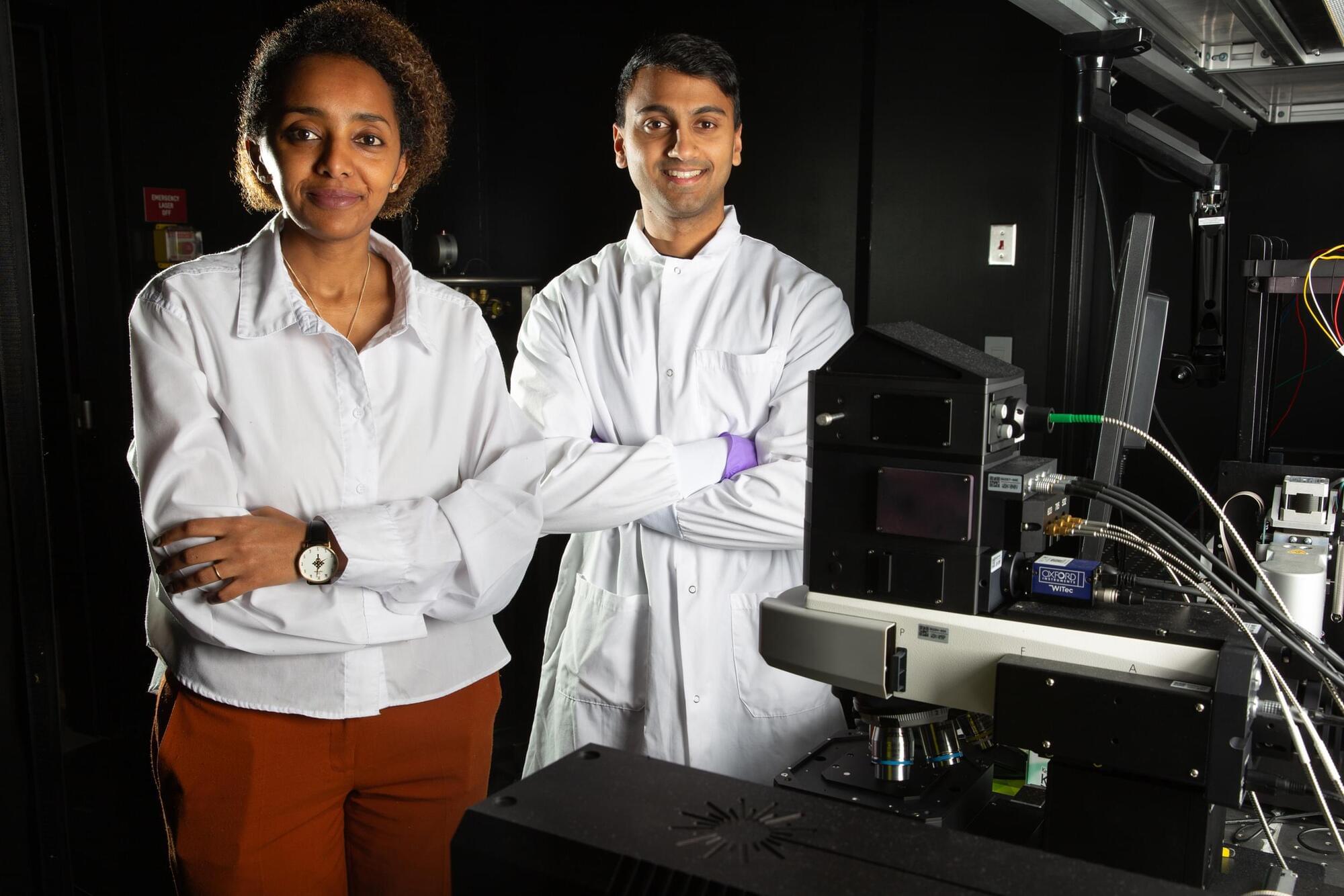
The researchers confirmed this by designing experiments that removed angular momentum while preserving helicity. The sideways rotation still occurred, showing that helicity plays the key role.
This finding offers a deeper understanding of how light interacts with matter at extremely small scales. It also points to new ways of controlling nanoscale systems, with possible applications in light-driven nanomachines and advanced sensing technologies.
“This work represents a new measurement paradigm for nanoscale optomechanics,” says Tanaka. “Just as optical tweezers opened a new field in single-molecule biophysics, we hope this platform will unlock access to nanoscale mechanical phenomena that have so far remained beyond reach.”
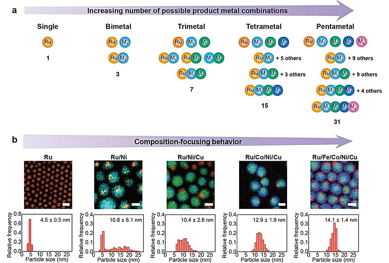
A nanocrystal is an extraordinarily tiny piece of material—composed of anywhere from a few to a few thousand atoms—in which atoms are arranged in a precise, ordered structure. Think of it like taking a piece of gold and shrinking it down to the size of a few hundred atoms. It’s still gold, still crystalline, just almost incomprehensibly small.
Nanocrystals are in the transistors inside computers and smartphones, in smartphone displays and TV screens, in the gold-nanoparticle sensors that power COVID and pregnancy tests, and in the pipes of your car exhaust system, among countless other innovations.
Their small size gives them a dramatically higher ratio of surface area to volume, making them especially useful as catalysts—materials that speed up chemical reactions without being consumed in the process.
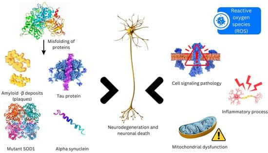
Neurodegenerative disorders entail a progressive loss of neurons in cerebral and peripheral tissues, coupled with the aggregation of proteins exhibiting altered physicochemical properties. Crucial to these conditions is the gradual degradation of the central nervous system, manifesting as impairments in mobility, aberrant behaviors, and cognitive deficits. Mechanisms such as proteotoxic stress, neuroinflammation, oxidative stress, and programmed cell death contribute to the ongoing dysfunction and demise of neurons. Presently, neurodegenerative diseases lack definitive cures, and available therapies primarily offer palliative relief. The integration of nanotechnology into medical practices has significantly augmented both treatment efficacy and diagnostic capabilities.
*** This content was analyzed and written by AI for informational purposes only. *** Please consult a specialist for professional advice.
The world is entering an era where “technology” and “living organisms” merge into one. Most recently, in 2026, a research team from Northwestern University created a landmark breakthrough by developing “Printed Neurons.” These are not designed just to mimic biology—they can actually “transmit signals” to communicate with living brain cells!
Why is this a big deal? Typically, the silicon-based computers we use today operate entirely differently from the human brain. Computers consume massive amounts of power and are rigid. In contrast, our brains use only about 20 watts (less than some lightbulbs) and are incredibly flexible. Creating artificial neurons that “speak the same language as the brain” is the key to treating diseases that were once considered incurable.
Innovations in “Electronic Ink” and “3D Printing“ At the heart of this research lies a leap forward in materials science and engineering: • Nanomaterials (MoS₂ and Graphene): Researchers used these materials to create a specialized “ink” for printing neural networks. These materials are unique for being both flexible and excellent conductors of electricity. • Aerosol Jet Printing: This technology allows for nano-level precision printing on flexible plastic sheets, designed to contour perfectly to human tissue. • Biomimicry: These artificial cells can generate electrical signals called “Spikes,” matching the rhythm and speed of actual biological neurons.
Proven! Successful Communication with a “Mouse Brain“ The research team tested the connection between these printed neurons and mouse brain tissue. The results showed that the mouse brain cells could receive and respond to signals from the artificial device as if they were from their own kind. This is vital evidence that humans can create devices that interface seamlessly with the nervous system.
Printed artificial neurons reported by Northwestern University can produce neuron-like electrical spikes and trigger responses in living mouse brain tissue. This video explains what was shown, why it matters for brain-like computing and future neural interfaces, and why it is still early laboratory research, not a human implant.
Scientists may have found a powerful new way to lower “bad” cholesterol, which did not involve the use of statin medicines. In a recent study, researchers used tiny DNA-based molecules to cut levels of LDL (bad) cholesterol by nearly 50% in animal models. This was done without the side effects that are often linked to statins. If these results stay consistent in large human trials, the therapy could become an important option for people who cannot tolerate statins or who still have high cholesterol despite taking them. The study was led by Carles J. Ciudad and Veronica Noe from the University of Barcelona’s Faculty of Pharmacy and Food Sciences and the Institute of Nanoscience and Nanotechnology (IN2UB), working with Nathalie Pamir at the University of Oregon in Portland (United States). It was published in the journal Biochemical Pharmacology.
High LDL cholesterol is one of the biggest risk factors for heart attacks and strokes because it leads to the build up fatty plaques in arteries. Drugs like statins work well for many, but some people suffer from muscle aches, digestive issues, or liver problems and have to stop them. However, the new approach is different. Instead of changing how the liver handles fats, it targets a specific protein in the blood that controls how much LDL stays circulating.
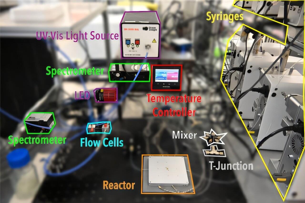
A new autonomous laboratory recently navigated through billions of potential material synthesis recipes to identify brighter, lead-free light-emitting nanomaterials in just 12 hours. The work could accelerate development of safer light-emitting nanoplatelets for use in applications ranging from photodetectors to the production of fuel from solar energy. A paper describing this work appears in Nature Communications.
Nanoplatelets are sheet-like crystals only billionths of a meter thick; in this case, they belong to a family of lead-free “double perovskites,” materials whose atomic recipe can be tuned to control how they absorb and emit light.
“One of the big challenges in developing safer optical nanomaterials is the sheer size of the material universe,” says Milad Abolhasani, Alcoa Professor and University Faculty Scholar in the department of chemical and biomolecular engineering at North Carolina State University. Abolhasani is the corresponding author of the research.
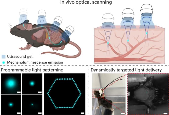
Beautifully executed paper on putting mechanoluminescent nanoparticles into blood circulation of mice which express optogenetic channels. Focused ultrasound can then trigger targeted light emission and control of neural activity in the brain and elsewhere.
A deep-tissue light source made from mechanoluminescent transducers stimulated by focused ultrasound enables wide imaging of live animal vasculature, and modulation of neuronal activity and behaviour.
Diagnosing some diseases could be as easy as breathing into a tube. MIT engineers have developed a test to detect disease-related compounds in a patient’s breath. The new test could provide a faster way to diagnose pneumonia and other lung conditions. Rather than sit for a chest X-ray or wait hours for a lab result, a patient may one day take a breath test and get a diagnosis within minutes.
The new breath test is a portable, chip-scale sensor that traps and detects synthetic compounds, or “biomarkers,” of disease, which are initially attached to inhalable nanoparticles. The biomarkers serve as tiny tags that can only be unlocked and detached from the nanoparticle by a very particular key, such as a disease-related enzyme.
The idea is that a person would first breathe in the nanoparticles, similar to inhaling asthma medicine. If the person is healthy, the nanoparticles would eventually circulate out of the body intact. If a disease such as pneumonia is present, however, enzymes produced as a result of the infection would snip off the nanoparticles’ biomarkers. These untethered biomarkers would be exhaled and measured, confirming the presence of the disease.
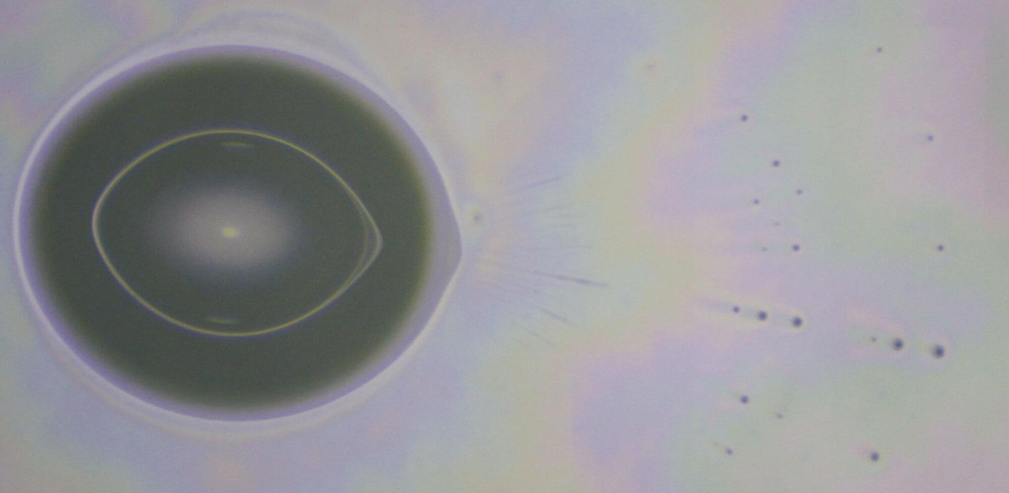
Water droplets might seem simple at first. But when nearing evaporation, a desperate power struggle of competing physical forces can emerge, with explosive effects. In a Proceedings of the National Academy of Sciences publication, researchers have taken a closer look at the physics of charged water droplets on frictionless surfaces, observing spontaneous jets of microdroplet emissions. Their insights may open new opportunities in nanoscale fabrication and electrospray ionization.
Professor Dan Daniel, head of the Droplet and Soft Matter Unit at the Okinawa Institute of Science and Technology (OIST) says, “From raindrops to spray coatings, mass spectrometry to microfluidics, sneezes to spacecraft plumes, charged droplets can show up in a surprising wealth of settings. Our observations enable new physical understanding of evaporating charged droplets, with a range of potential industrial applications.”