What it means to be conscious is more than just a philosophical question. Researchers continue to investigate how conscious experience arises from the electrochemical activity of the human brain. The answer has important implications for the way brain health is understood—from coma, wherein a person is alive but unable to move or respond to his or her environment, to surgical anesthesia, to the altered thought processes of schizophrenia.
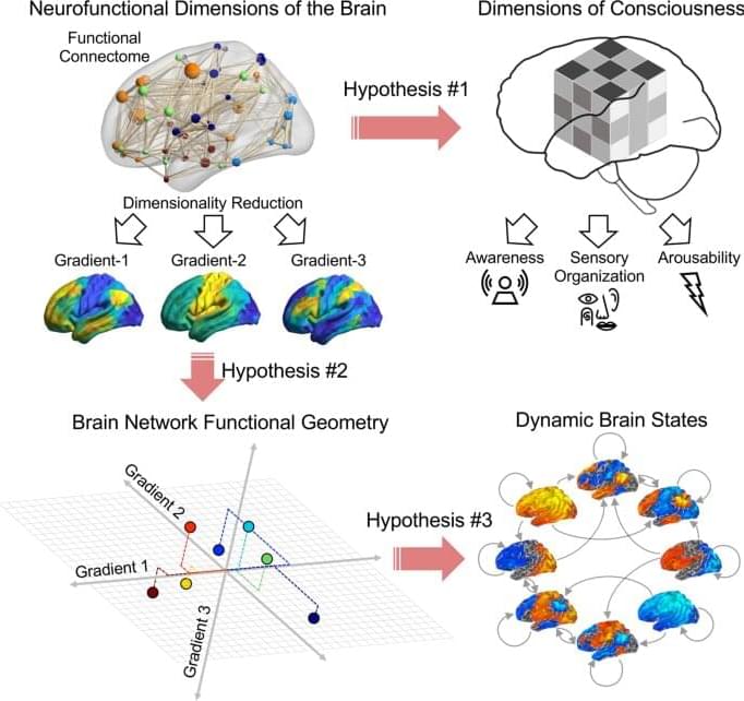
Recent research suggests that there’s no one location in the brain that causes consciousness, pointing to a network phenomenon. However, tracing the various linkages between regions in the brain networks that give rise to awareness and wakefulness has been elusive.
A new approach using functional MRI, an imaging technique that allows you to see and measure brain activity through changes in blood flow over time, provides new insight into how we describe and study conscious states.