Toggle light / dark theme

Laser plasma accelerator achieves enhanced electron beam quality for practical applications

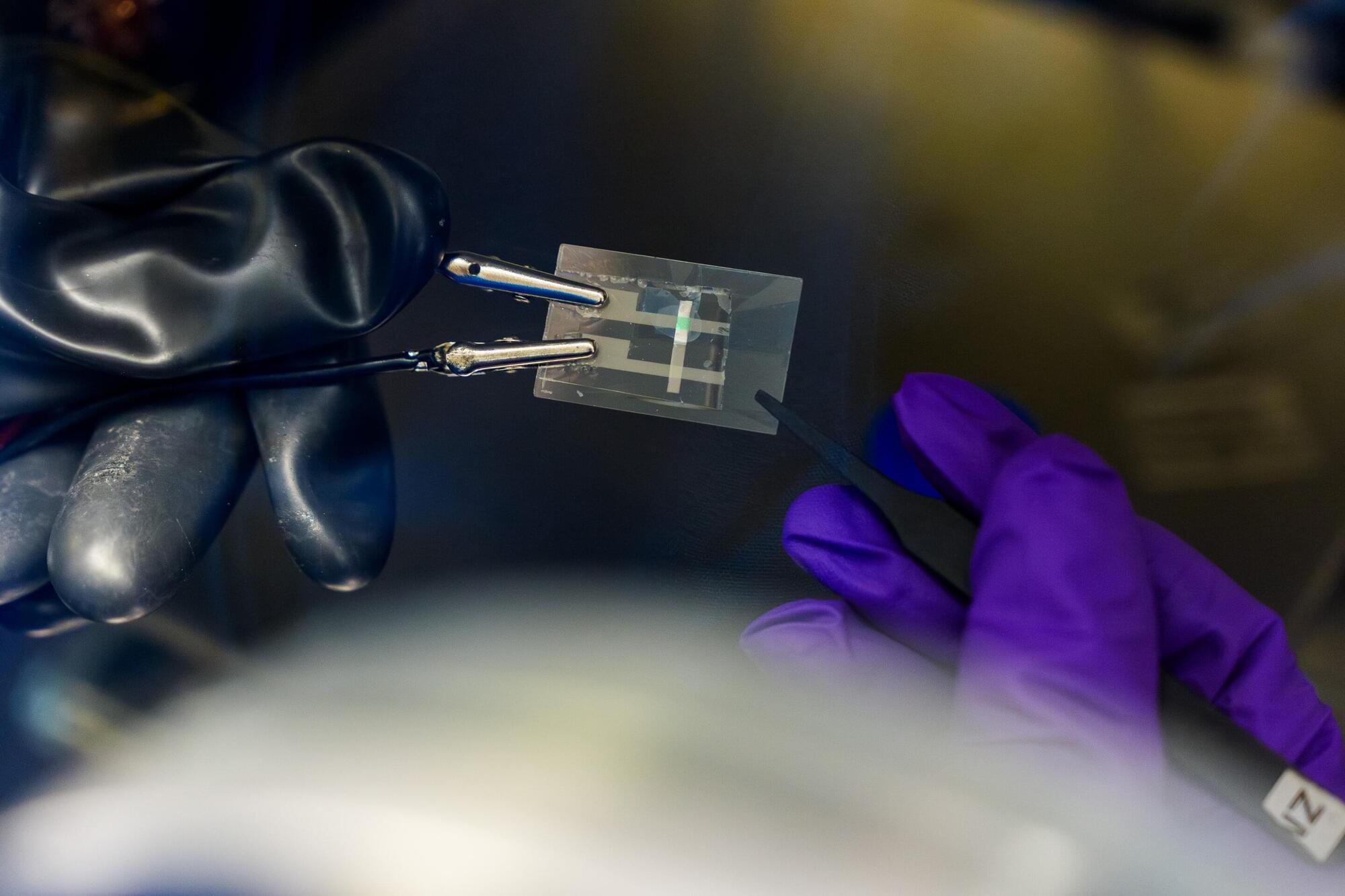
Laser plasma acceleration is a potentially disruptive technology: It could be used to build far more compact accelerators and open up new use cases in fundamental research, industry and health. However, on the path to real-world applications, some properties of the plasma-driven electron beam as delivered by current prototype accelerators still need to be refined.

DESY’s LUX experiment has now made significant progress in this direction: Using a clever correction system, a research team was able to significantly improve the quality of electron bunches accelerated by a laser plasma accelerator. This brings the technology a step closer to concrete applications, such as a plasma-based injector for a synchrotron storage ring. The research group presents their results in the journal Nature.

Conventional electron accelerators use which are directed into so-called resonator cavities. The radio waves transfer energy to the electrons as they fly past, increasing their velocity. To achieve high energies, many resonators have to be connected in series, making the machines large and costly.

A Smart Wearable for Ear-Based High-Precision Health Sensing

Wearables such as smartwatches, fitness trackers, or data glasses have become an integral part of our everyday lives. They record health data, monitor your sleep, or calculate your calorie consumption. Researchers from Karlsruhe Institute of Technology (KIT) have developed the open-source platform “OpenEarable.” It integrates a multitude of sensors into wireless earphones with the aim to enhance health measurements and safety applications in medicine, industry, and everyday life. The scientists are currently presenting their platform at Hannover Messe from March 31 to April 4.

Wearable technologies have made significant progress in recent years, but many of the existing systems are either proprietary, i.e. not customizable by others, or their measurement capabilities are limited. With OpenEarable 2.0, a research team headed by Dr. Tobias Röddiger from KIT’s TECO research group moves one step further: The open-source platform for ear-based sensor applications enables developers to create customized software. With a unique combination of sensors, more than 30 physiological parameters can be measured directly at the ear – from heart rate and breathing patterns to fatigue and body temperature. “Our aim was to create an open and high-precision solution for health monitoring that goes far beyond what is possible with today’s commercial wearables,” says Röddiger. “OpenEarable 2.0 provides a platform for researchers and developers that is easily customizable and scalable. This allows them to program the earphones individually for specific requirements.

MicroRNA regulation of enteric nervous system development and disease

MicroRNAs (miRNAs) serve as key regulators of enteric nervous system development, orchestrating migration, proliferation, and differentiation of enteric nervous system progenitors.

Aberrant miRNA expression underpins the pathogenesis of several enteric neuropathies, including Hirschsprung’s disease.

A convergence of miRNA activity across distinct enteric neuropathies highlights shared molecular pathways, exemplified by the miR-200 family.

Modulating the expression of miRNAs to influence their associated gene expression networks has therapeutic potential for enteric neuropathies. https://sciencemission.com/MicroRNA-regulation-of-enteric-ne…nd-disease


The enteric nervous system (ENS), an elaborate network of neurons and glia woven through the gastrointestinal tract, is integral for digestive physiology and broader human health. Commensurate with its importance, ENS dysfunction is linked to a range of debilitating gastrointestinal disorders. MicroRNAs (miRNAs), with their pleiotropic roles in post-transcriptional gene regulation, serve as key developmental effectors within the ENS. Herein, we review the regulatory dynamics of miRNAs in ENS ontogeny, showcasing specific miRNAs implicated in both congenital and acquired enteric neuropathies, such as Hirschsprung’s disease (HSCR), achalasia, intestinal neuronal dysplasia (IND), chronic intestinal pseudo-obstruction (CIPO), and slow transit constipation (STC).

AI could revolutionize early skin cancer detection, treatment

Health care providers can use small devices to hover over moles or lesions and immediately check for common skin cancers, such as melanoma and basal cell carcinoma.

The most significant benefit is that health care professionals who do not specialize in dermatology could perform these checks during a routine visit, making early detection easier and quicker.

Skin cancer is the most common form of cancer in the United States, with one in five Americans expected to be affected in their lifetime, according to the City of Hope Cancer Center.

From social to biological networks: New algorithm uncovers key proteins in human disease

Researchers at Ben-Gurion University of the Negev have developed a machine-learning algorithm that could enhance our understanding of human biology and disease. The new method, Weighted Graph Anomalous Node Detection (WGAND), takes inspiration from social network analysis and is designed to identify proteins with significant roles in various human tissues.

Proteins are essential molecules in our bodies, and they interact with each other in , known as (PPI) networks. Studying these networks helps scientists understand how proteins function and how they contribute to health and disease.

Prof. Esti Yeger-Lotem, Dr. Michael Fire, Dr. Jubran Juman, and Dr. Dima Kagan developed the algorithm to analyze these PPI networks to detect “anomalous” proteins—those that stand out due to their unique pattern of weighted interactions. This implies that the amount of the protein and its protein interactors is greater in that particular network, allowing them to carry out more functions and drive more processes. This also indicates the great importance that these proteins have in a particular network, because the body will not waste energy on their production for no reason.

Your season of conception could influence how your body stores fat

Individuals who were conceived in colder seasons are more likely to show higher brown adipose tissue activity, increased energy expenditure and a lower body mass index (BMI), and lower fat accumulation around internal organs, compared with those conceived in warmer seasons, suggests a study published in Nature Metabolism. The findings, based on an analysis involving more than 500 participants, indicate a potential role for meteorological conditions influencing human physiology.

Although eating habits and exercise are key indicators of fat loss, exposure to cold and warmth also plays a part. In colder temperatures, the body generates more heat (cold-induced thermogenesis) via brown adipose tissue activity and stores less fat in the form of white adipose tissue than it does in hotter temperatures. However, underlying factors contributing to in brown adipose tissue activity remain poorly understood, particularly in humans.

Takeshi Yoneshiro and colleagues analyzed brown adipose tissue density, activity and thermogenesis in 683 healthy male and female individuals between ages 3 and 78 in Japan, whose parents were exposed to (defined in the study as between 17 October and 15 April) or warm temperatures (between 16 April and 16 October) during the fertilization and birth periods.

Abstract: This work was supported in part by the NIH National Cancer Institute grants R01 CA186338

R01 CA203108, R01 CA247234 (to ML), and by the William and Ella Owens Medical Research Foundation (to ML). It was also supported in part by the Department of Medicine, the University of Oklahoma Health Sciences Center.

Address correspondence to: Michael S. Bronze, Department of Medicine, The University of Oklahoma Health Sciences Center, 800 Stanton L. Young Blvd. AAT 6,400, Oklahoma City, Oklahoma, 73,104, USA. Phone: 405.271.5428; Email: [email protected]. Or to: Min Li, Department of Medicine, The University of Oklahoma Health Sciences Center, 975 NE 10th Street, BRC 1262A, Oklahoma City, Oklahoma, 73,104, USA. Phone: 405.271.1796; Email: [email protected].