Join us on Patreon! https://www.patreon.com/MichaelLustgartenPhDDiscount Links: Epigenetic Testing: https://trudiagnostic.com/?irclickid=U-s3Ii2r7xyIU-LSYLyQ…
Category: genetics – Page 210

Johns Hopkins Scientists Solve 30-Year Biological Mystery of Night Blindness
In what they believe is a solution to a 30-year biological mystery, neuroscientists at Johns Hopkins Medicine say they have used genetically engineered mice to address how one mutation in the gene for the light-sensing protein rhodopsin results in congenital stationary night blindness.
The condition, present from birth, causes poor vision in low-light settings.
The findings, published May 14 in Proceedings of the National Academy of Sciences, demonstrate that the rhodopsin gene mutation, called G90D, produces an unusual background electrical “noise” that desensitizes the eye’s rods, those cells in the retina at the back of the eye responsible for nighttime vision, thus causing night blindness.

Combining proteomics and AI to enable ‘a new era in healthcare’
Understanding aging and age-related diseases requires analyzing a vast number of factors, including an individual’s genetics, immune system, epigenetics, environment and beyond. While AI has long been touted for its potential to shed light on these complexities of human biology and enable the next generation of healthcare, we’ve yet to see the emergence of tools that truly deliver on this promise.
Leveraging advanced plasma proteomics, US startup Alden Scientific has developed AI models capable of making the connections needed to accurately assess an individual’s state of health and risk of disease. The company’s tool measures more than 200 different conditions, including leading causes of morbidity and mortality such as Alzheimer’s, heart disease, diabetes and stroke. Significantly, its models also enable an individual to understand how an intervention impacts these risks.
With a host of top Silicon Valley investors among its early adopters, Alden is now using its platform to conduct an IRB-approved health study designed to provide a “longitudinal understanding of the interplay between environmental, biological, and medical data.”
A Neurodevelopmental Disorder Affecting Thousands is Discovered
For decades, the study of genetic disease was focused on genes that code for protein. But scientists have now identified a novel neurodevelopmental disorder that is caused by mutations in a gene that does not code for protein, called RNU4-2. These mutations lead to neurological symptoms that cause cognitive dysfunction, but have not previously been linked together as one disease. These findings have been reported in Nature Medicine.
In this work, the investigators analyzed whole-genome sequencing data from over 5,000 cases of intellectual disability and over 46,000 unaffected individuals. The research focused on unusual variations in the sequences of 41,132 genes that do not code for protein. The research revealed a gene that is apparently a common cause of neurodevleopmental problems. The scientists also determined that these mutations often arise spontaneously, and are not usually inherited from a parent.
Editing without ‘cutting’: Molecular mechanisms of new gene-editing tool revealed
Joint research led by Yutaro Shuto, Ryoya Nakagawa, and Osamu Nureki of the University of Tokyo determined the spatial structure of various processes of a novel gene-editing tool called “prime editor.” Functional analysis based on these structures also revealed how a “prime editor” could achieve reverse transcription, synthesizing DNA from RNA, without “cutting” both strands of the double helix. Clarifying these molecular mechanisms contributes greatly to designing gene-editing tools accurate enough for gene therapy treatments. The findings were published in the journal Nature.
The 2020 Nobel Prize in Chemistry was awarded to Jennifer Doudna and Emmanuelle Charpentier for developing a groundbreaking yet simple way to edit DNA, the “blueprint” of living organisms. While their discovery opened new avenues for research, the accuracy of the method and safety concerns about “cutting” both strands of DNA limited its use for gene therapy treatments. As such, research has been underway to develop tools that do not have these drawbacks.
The prime editing system is one such tool, a molecule complex consisting of two components. One component is the prime editor, which combines a SpCas9 protein, used in the first CRISPR-Cas gene editing technology, and a reverse transcriptase, an enzyme that transcribes RNA into DNA. The second component is the prime editing guide RNA (pegRNA), a modified guide RNA that identifies the target sequence within the DNA and encodes the desired edit. In this complex, the prime editor works like a “word processor,” accurately replacing genomic information. The tool has already been successfully implemented in living cells of organisms such as plants, zebrafish, and mice. However, precisely how this molecule complex executes each step of the editing process has not been clear, mostly due to a lack of information on its spatial structure.
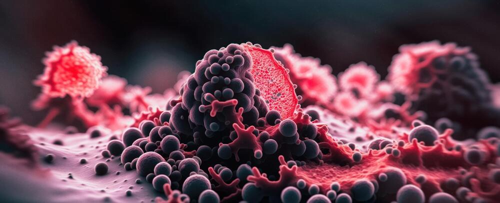
Reversible Molecular Changes Can Cause Cancer, Study Shows
Though one in two people will develop some form of cancer in their lifetime, there’s still much we don’t know about this disease. But thanks to continued research efforts, we keep learning more about the biology of cancer. One of these recent discoveries could even transform our understanding of how cancers develop.
But before we talk about the new discovery, let’s first discuss the classical theory that attempts to explain why normal cells become cancer cells. This theory posits that DNA mutations are the primary cause of cancers.
It’s well known that ageing, as well as some lifestyle and environmental factors (such as smoking and UV radiation) cause random DNA mutations (also known as genetic alterations) in our cells. Most genetic alterations trigger cell death or have no consequence.

Maximizing DNA Yield for Biobanking Applications
With advances in genomics research, personalized medicine, and sequencing-based technologies, there is a necessity for purification of high-quality genomic DNA from large volumes of blood. The rapidly growing landscape of biorepositories that store large amounts of DNA from an enormous number of biospecimens further fuels this need to find optimized solutions for reliable purification of DNA. The information derived from the purified DNA is crucial to health science research and facilitates drug discovery, biomarker discovery, clinical implementation projects, etc. For the success of these analyses and to derive relevant information, DNA extraction is the most critical step and must meet the criteria of extraction speed, yield and quality, as well as reproducibility. Many nucleic acid purification kits and automation workflows for processing blood samples in the volume range of 100–250 μL exist, but not many convenient, automated options exist for volumes as high as 2 mL without sample splitting. To fill this opening, Omega Bio-tek has developed a semi-automated solution on the MagBinder® Fit24 to extract DNA from large volumes of fresh or frozen blood. Here, we provide background information on biobanks, as well as present the solution Omega Bio-tek has developed for DNA extraction from large volumes of whole blood.
A biobank is a specialized repository that systematically collects, processes, stores, and manages biological samples for use in medical research and treatments. The primary purpose of a biobank is to provide a centralized and organized resource of high-quality biological materials, such as blood or tissue, along with relevant clinical and demographic data1. These invaluable assets are at the center of advancements in cancer treatments, biomarker discovery, and understanding genetic factors for disease. At a high level, biobanks can be classified by two categories1:
Upending Assumptions About the Uniformity of DNA in the Human Body
It’s long been assumed that as cells divide in the human body, the genome is faithfully replicated in the resulting daughter cells. While errors are known to arise, there is machinery in the cell that can detect these genetic errors, and can often repair them. When mutations remain in the genome, it raises the risk that disease will arise.
But the human genome is made up of about 6 billion bases, and the human body contains billions, even trillions of cells. And it seems that errors and variations in the genome could actually arise far more often than we knew, according to a new study reported in Nature Genetics that analyzed blood stem cells. The research used advanced sequencing techniques to show that humans are made up of cells whose genomes may be far more heterogeneous that assumed. And these variations between cells are not always small. The research determined that about one out of every forty blood stem cells in healthy people carry major chromosomal alterations in their DNA. These chromosomal changes included copy number variations and rearrangements, but did not seem to cause any deleterious effect.

Scientists Pinpoint Main Cause of Sensory Hypersensitivity in Autism
Sensory hypersensitivity in mice with the Grin2b gene mutation found in patients is related to hyperactivity of the anterior cingulate cortex (ACC) and hyperconnectivity between the ACC and other brain regions. Credit: Institute for Basic Science.
Director Kim Eunjoon states, “This new research demonstrates the involvement of the anterior cingulate cortex (ACC), which has been known for its deep association with cognitive and social functions, in sensory hypersensitivity in autism.”
The hyperactivity of the ACC was also associated with the enhanced functional connectivity between the ACC and other brain areas. It is believed both hyperactivity and the hyperconnectivity of the ACC with various other brain regions are involved with sensory hypersensitivity in Grin2b-mutant mice.