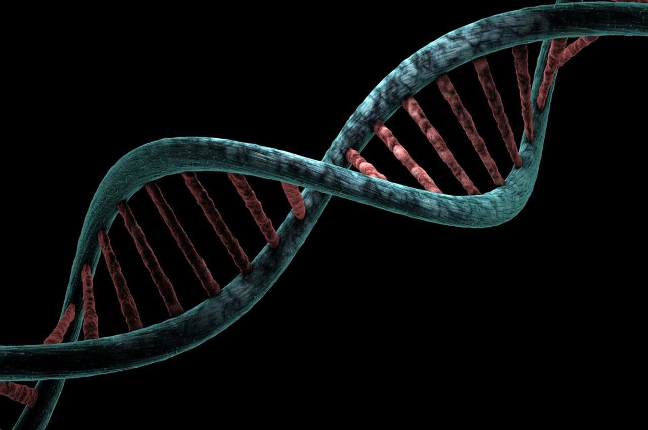
Toggle light / dark theme

Study Links Gene Variations to Brain Changes in Essential Tremor

Summary: Researchers identified how gene variations lead to brain changes associated with essential tremor, a common movement disorder affecting over 60 million people worldwide. The study used brain MRI scans and genetic data from over 33,000 adults to uncover genetic links to structural changes in the brain’s cortex and cerebellum.

These findings could lead to new drug targets by revealing how faulty protein disposal systems disrupt neural pathways, resulting in uncontrollable hand tremors. The research marks a significant step toward understanding and treating essential tremor more effectively.

Nucleated synthetic cells with genetically driven intercompartment communication

The brain’s white matter comprises areas of the central nervous system made up of myelinated axons. Its name is derived from the pale appearance of the lipids that comprise myelin. Myelin is a segmented sheath that insulates axons, ensuring the conduction of neural signals. The loss of myelin is documented in a number of neurodegenerative pathologies, including Alzheimer’s and Parkinson’s disease, and perhaps most notably, multiple sclerosis. As people age, demyelination becomes more likely.

Researchers have long suspected a relationship between and the integrity of the brain’s as people age. However, a lack of specific evidence has led researchers at the National Institutes of Health to conduct a study examining the strength of this correlation, now published in the Proceedings of the National Academy of Sciences.

To establish a correlation between cardiovascular fitness and cerebral myelination, the researchers recruited a cohort of 125 participants from age 22 to 94 years old. The cardiovascular fitness of the participants was quantified as the maximum rate of oxygen consumption, popularly and succinctly known as VO2max. Myelin content was defined as the water fraction, which the researchers estimated through an advanced multicomponent relaxometry MRI method.

Collaborative research cracks the autism code, making the neurodivergent brain visible

A multi-university research team co-led by University of Virginia engineering professor Gustavo K. Rohde has developed a system that can spot genetic markers of autism in brain images with 89 to 95% accuracy.

Their findings suggest that doctors may one day see, classify and treat autism and related neurological conditions with this method, without having to rely on or wait for behavioral cues. And that means this truly personalized medicine could result in earlier interventions.

“Autism is traditionally diagnosed behaviorally but has a strong genetic basis. A genetics-first approach could transform understanding and treatment of autism,” the researchers wrote in a paper published in the journal Science Advances.

Non-cognitive skills: DNA-based analyses suggest a hidden key to academic success

A new Nature Human Behaviour study, jointly led by Dr. Margherita Malanchini at Queen Mary University of London and Dr. Andrea Allegrini at University College London, has revealed that non-cognitive skills, such as motivation and self-regulation, are as important as intelligence in determining academic success. These skills become increasingly influential throughout a child’s education, with genetic factors playing a significant role.

The research, conducted in collaboration with an international team of experts, suggests that fostering non-cognitive skills alongside could significantly improve educational outcomes.

“Our research challenges the long-held assumption that intelligence is the primary driver of ,” says Dr. Malanchini, Senior Lecturer in Psychology at Queen Mary University of London.

Scientists discover new code governing gene activity

A newly discovered code within DNA—coined “spatial grammar”—holds a key to understanding how gene activity is encoded in the human genome.

This breakthrough finding, identified by researchers at Washington State University and the University of California, San Diego and published in Nature, revealed a long-postulated hidden spatial grammar embedded in DNA. The research could reshape scientists’ understanding of and how genetic variations may influence in development or disease.

Transcription factors, the proteins that control which genes in one’s genome are turned on or off, play a crucial role in this code. Long thought of as either or repressors of gene activity, this research shows the function of transcription factors is far more complex.

Scientists Discover “Spatial Grammar” in DNA: Breakthrough Could Rewrite Genetics Textbooks

Researchers have discovered a “spatial grammar” in DNA that redefines the role of transcription factors in gene regulation, influencing our understanding of genetic variations and disease.

A recently uncovered code within DNA, referred to as “spatial grammar,” may unlock the secret to how gene activity is encoded in the human genome.

This breakthrough finding, identified by researchers at Washington State University and the University of California, San Diego and published in Nature, revealed a long-postulated hidden spatial grammar embedded in DNA. The research could reshape scientists’ understanding of gene regulation and how genetic variations may influence gene expression in development or disease.

Frontiers: Brain organoids have become increasingly used systems allowing 3D-modeling of human brain development, evolution, and disease

To be able to make full use of these modeling systems, researchers have developed a growing toolkit of genetic modification techniques. These techniques can be applied to mature brain organoids or to the preceding embryoid bodies (EBs) and founding cells. This review will describe techniques used for transient and stable genetic modification of brain organoids and discuss their current use and respective advantages and disadvantages. Transient approaches include adeno-associated virus (AAV) and electroporation-based techniques, whereas stable genetic modification approaches make use of lentivirus (including viral stamping), transposon and CRISPR/Cas9 systems. Finally, an outlook as to likely future developments and applications regarding genetic modifications of brain organoids will be presented.

The development of brain organoids (Kadoshima et al., 2013; Lancaster et al., 2013) has opened up new ways to study brain development and evolution as well as neurodevelopmental disorders. Brain organoids are multicellular 3D structures that mimic certain aspects of the cytoarchitecture and cell-type composition of certain brain regions over a particular developmental time window (Heide et al., 2018). These structures are generated by differentiation of induced pluripotent stem cells (iPSCs) or embryonic stem cells (ESCs) into embryoid bodies followed by, or combined, with neural induction (Kadoshima et al., 2013; Lancaster et al., 2013). In principle, two different classes of brain organoid protocols can be distinguished, namely: (i) the self-patterning protocols which produce whole-brain organoids; and (ii) the pre-patterning protocols which produce brain region-specific organoids (Heide et al., 2018).

Scientists devise Method to Secure Earth’s Biodiversity on the Moon

Proposed lunar biorepository could store genetic samples without electricity or liquid nitrogen. New research led by scientists at the Smithsonian proposes a plan to safeguard Earth’s imperiled biodiversity by cryogenically preserving biological material on the moon. The moon’s permanently shadowed craters are cold enough for cryogenic preservation without the need for electricity or liquid nitrogen, according to the researchers.

The paper, published today in BioScience and written in collaboration with researchers from the Smithsonian’s National Zoo and Conservation Biology Institute (NZCBI), Smithsonian’s National Museum of Natural History, Smithsonian’s National Air and Space Museum and others, outlines a roadmap to create a lunar biorepository, including ideas for governance, the types of biological material to be stored and a plan for experiments to understand and address challenges such as radiation and microgravity. The study also demonstrates the successful cryopreservation of skin samples from a fish, which are now stored at the National Museum of Natural History.

“Initially, a lunar biorepository would target the most at-risk species on Earth today, but our ultimate goal would be to cryopreserve most species on Earth,” said Mary Hagedorn, a research cryobiologist at NZCBI and lead author of the paper. “We hope that by sharing our vision, our group can find additional partners to expand the conversation, discuss threats and opportunities and conduct the necessary research and testing to make this biorepository a reality.”

/* */