Humanity’s future is now framed by artificial intelligence (AI) and increasing interventions in the human body to cure and to enhance, i.e., transhumanism. Considering the stakes and the dangers, a philosophical perspective is imperative.
Make a tax-deductible donation of any amount to help support Closer To Truth continue making content like this: https://shorturl.at/OnyRq.
Terrence William Deacon is an American neuroanthropologist. He taught at Harvard for eight years, relocated to Boston University in 1992, and is currently Professor of Anthropology and member of the Cognitive Science Faculty at the University of California, Berkeley.
Paul Thagard is Distinguished Professor Emeritus of Philosophy at the University of Waterloo and a Fellow of the Royal Society of Canada, the Cognitive Science Society, and the Association for Psychological Science. His work focuses on cognitive science, philosophy of mind, and the philosophy of science and medicine.
A team of neuroscientists has uncovered evidence that genetic influences on intelligence may operate through the density of brain wiring, highlighting a potential biological bridge between inherited DNA differences and the brain structures that support reasoning and problem-solving.
Visionary, patient-centric health research for all — dr. julia moore vogel, phd — scripps research / long covid treatment trial.
Dr. Julia Moore Vogel, PhD, MBA is Assistant Professor and Senior Program Director at The Scripps Research Institute (https://www.scripps.edu/science-and-me… where she is responsible for managing a broad portfolio of patient-centric health research studies, including The Long COVID Treatment Trial (https://longcovid.scripps.edu/locitt-t/), a fully remote, randomized, placebo-controlled clinical trial targeting individuals with long COVID, testing whether the drug Tirzepatide can reduce or alleviate symptoms of long COVID.
Prior to this current role, Dr. Vogel managed The Participant Center (TPC) for the NIH All of Us Research Program (https://www.scripps.edu/science-and-me… which was charged with recruiting and retaining 350,000 individuals that represent the diversity of the United States. TPC aims to make it possible for interested individuals anywhere in the US to become active participants, for example by collaborating with numerous outreach partners to raise awareness, collecting biosamples nationwide, returning participants’ results and developing self-guided workflows that enable participants to join whenever is convenient for them.
Prior to joining the Scripps Research Translational Institute, Dr. Vogel created, proposed, fundraised for, and implemented research and clinical genomics initiatives at the New York Genome Center and The Rockefeller University. She oversaw the proposal and execution of grants, including a $44M NIH Center for Common Disease Genomics in collaboration with over 20 scientific contributors across seven institutions. She also managed corporate partnerships, including one with IBM that assessed the relative value of several genomic assays for cancer patients.
Dr. Vogel has a BS in Mathematics from Rensselaer Polytechnic Institute, a PhD in Computational Biology and Medicine from Cornell and an MBA from Cornell.
Polygenic risk scores estimate genetic predisposition to complex diseases by combining thousands of DNA variants. Dr. Anna CF Lewis of Harvard Medical School discusses their promise for personalized medicine and the ethical challenges they raise.
Mayo Clinic researchers have developed a new tool that can estimate a person’s risk of developing memory and thinking problems associated with Alzheimer’s disease years before symptoms appear.
The research, published in The Lancet Neurology, builds on decades of data from the Mayo Clinic Study of Aging—one of the world’s most comprehensive population-based studies of brain health.
The study found that women have a higher lifetime risk than men of developing dementia and mild cognitive impairment (MCI), a transitional stage between healthy aging and dementia that often affects quality of life but still allows people to live independently. Men and women with the common genetic variant, APOE ε4, also have a higher lifetime risk.
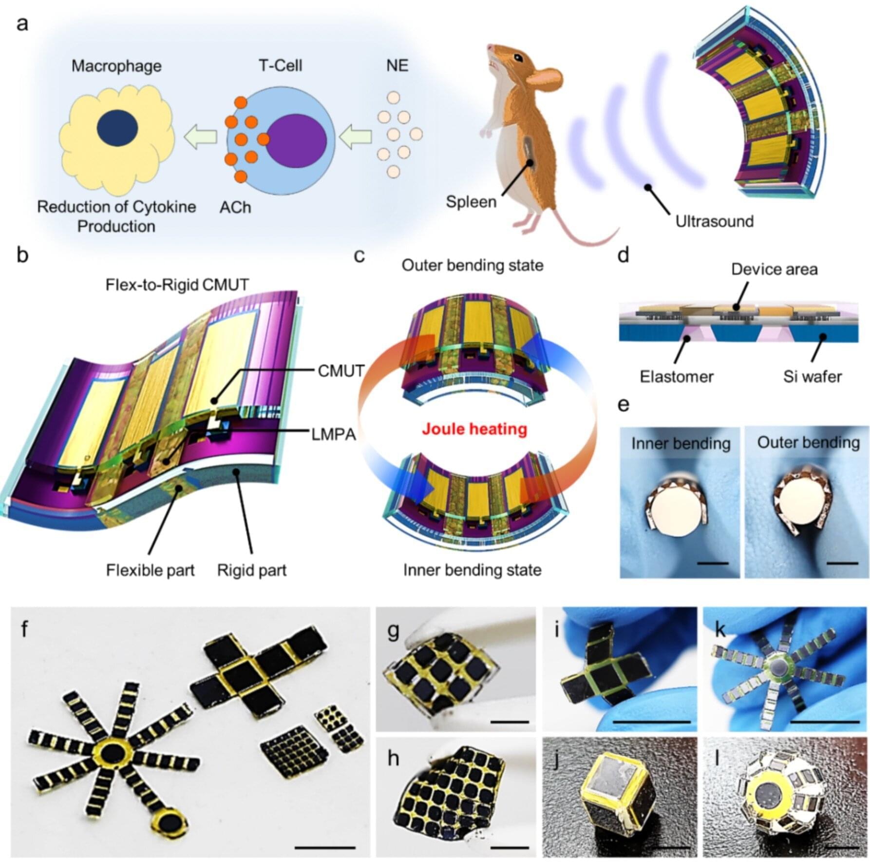
Conventional wearable ultrasound sensors have been limited by low power output and poor structural stability, making them unsuitable for high-resolution imaging or therapeutic applications.
A KAIST research team has now overcome these challenges by developing a flexible ultrasound sensor with statically adjustable curvature. This breakthrough opens new possibilities for wearable medical devices that can capture precise, body-conforming images and perform noninvasive treatments using ultrasound energy.
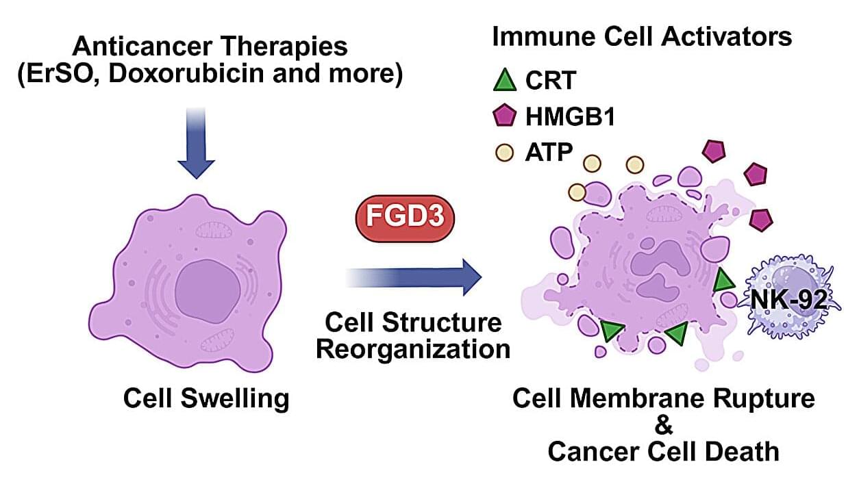
A naturally-occurring protein that tends to be expressed at higher levels in breast cancer cells boosts the effectiveness of some anticancer agents, including doxorubicin, one of the most widely used chemotherapies, and a preclinical drug known as ErSO, researchers report. The protein, FGD3, contributes to the rupture of cancer cells disrupted by these drugs, boosting their effectiveness and enhancing anticancer immunotherapies.
The discovery is described in the Journal of Experimental & Clinical Cancer Research.
The new findings were the result of experiments involving ErSO, an experimental drug that killed 95–100% of estrogen-receptor-positive breast cancer cells in a mouse model of the disease.
Dr Andrea Weisse, from the University of Edinburgh’s Schools of Biological Sciences and Informatics, who led the research, highlighted the urgency of the situation.
“Bacteria are clever little things. They have been learning how to dodge our antibiotics, and they are getting better at it all the time,” she said.
“If we don’t find new drugs – or new tricks to outsmart them – we are in trouble. What we are trying to do here is really understand how their defence systems work. Once we see the mechanism clearly, we can figure out smarter ways to beat them and treat infections more effectively.”