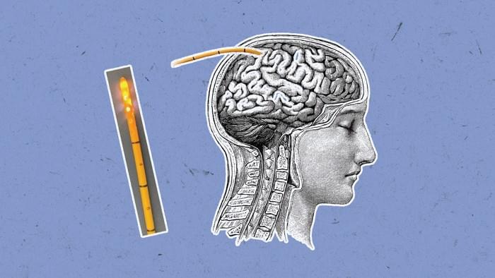
The new study demonstrates the ability to learn new skills without conscious effort.
Researchers have developed a groundbreaking technique using brain imaging and neurofeedback to induce learning without conscious effort.

Chronological age (CA) does not reflect individual variation in the aging process. However, existing biological age predictors are mostly based on European populations and overlook the widespread nonlinear effects of clinical biomarkers.
Using data from the prospective Dongfeng-Tongji (DFTJ) cohort of elderly Chinese, we propose a physiological aging index (PAI) based on 36 routine clinical biomarkers to measure aging progress. We first determined the optimal level of each biomarker by restricted cubic spline Cox models. For biomarkers with a U-shaped relationship with mortality, we derived new variables to model their distinct effects below and above the optimal levels. We defined PAI as a weighted sum of variables predictive of mortality selected by a LASSO Cox model. To measure aging acceleration, we defined ΔPAI as the residual of PAI after regressing on CA. We evaluated the predictive value of ΔPAI on cardiovascular diseases (CVD) in the DFTJ cohort, as well as nine major chronic diseases in the UK Biobank (UKB).
In the DFTJ training set (n = 12,769, median follow-up: 10.38 years), we identified 25 biomarkers with significant nonlinear associations with mortality, of which 11 showed insignificant linear associations. By incorporating nonlinear effects, we selected CA and 17 clinical biomarkers to calculate PAI. In the DFTJ testing set (n = 15,904, 5.87 years), PAI predict mortality with a concordance index (C-index) of 0.816 (95% confidence interval, [0.796, 0.837]), better than CA (C-index = 0.771 [0.755, 0.788]) and PhenoAge (0.799 [0.784, 0.814]). ΔPAI was predictive of incident CVD and its subtypes, independent of traditional risk factors. In the external validation set of UKB (n = 296,931, 12.80 years), PAI achieved a C-index of 0.749 (0.746, 0.752) to predict mortality, remaining better than CA (0.706 [0.702, 0.709]) and PhenoAge (0.743 [0.739, 0.746]).
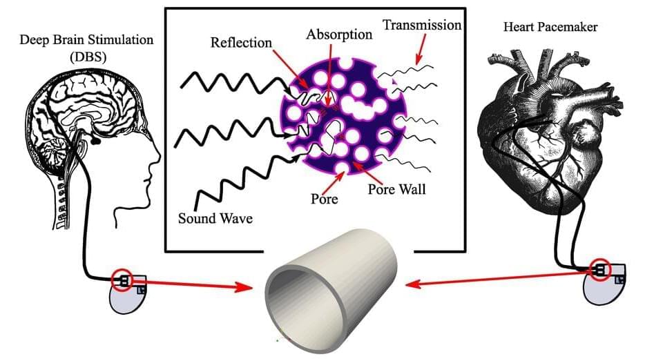
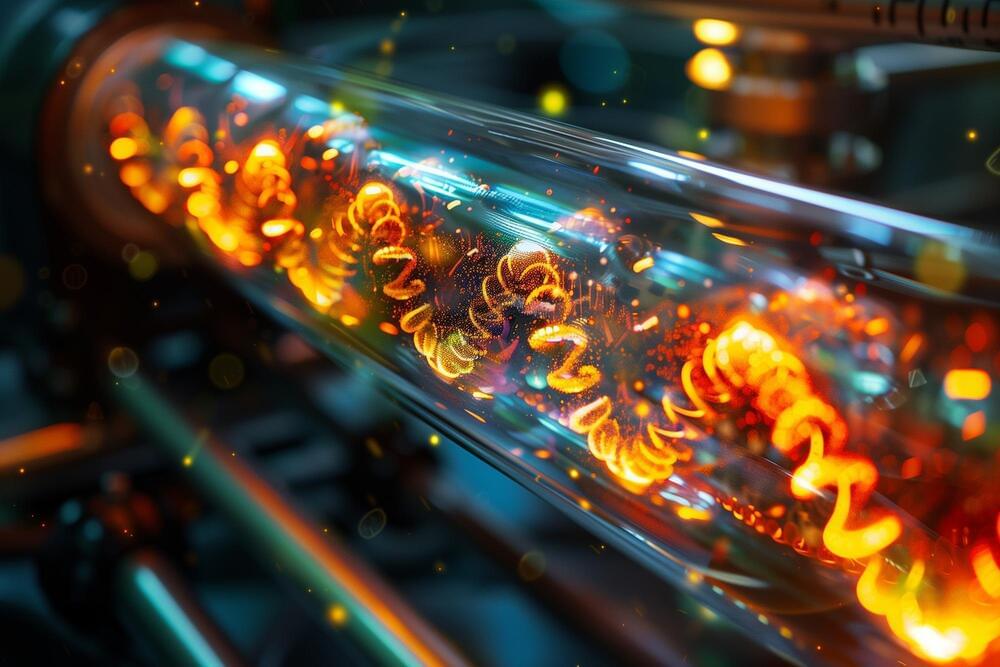
Two years ago, a medical professional approached scientists at the University of Tabriz in Iran with an interesting problem: Patients were having headaches after pacemaker implants. Working together to investigate, they began to wonder if the underlying issue is the materials used in the pacemakers.
“Managing external noise that affects patients is crucial,” author Baraa Chasib Mezher said. “For example, a person with a brain pacemaker may experience interference from external electrical fields from phones or the sounds of cars, as well as various electromagnetic forces present in daily life. It is essential to develop novel biomaterials for the outlet gate of brain pacemakers that can effectively handle electrical signals.”
In an article published this week in AIP Advances, Mezher, who is an Iraqi doctoral student studying in Iran, and her colleagues at the Nanostructured and Novel Materials Laboratory at the University of Tabriz created organic materials for brain and heart pacemakers, which rely on uninterrupted signal delivery to be effective.
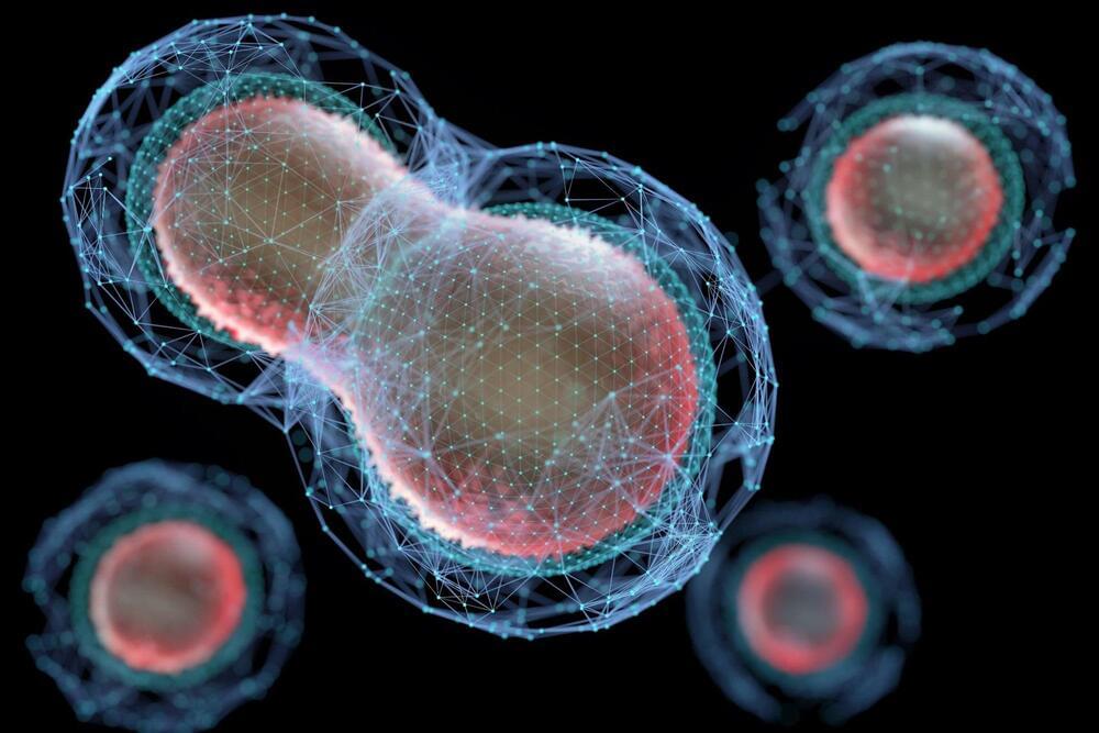
Cellular death is a fundamental concept in biological sciences. Despite its importance, its definition varies depending on the context in which it occurs and lacks a general mathematical definition.
Researchers from the University of Tokyo propose a new mathematical definition of death based on whether a potentially dead cell can return to a predefined “representative state of living,” which are the states of being that we can confidently call “alive.” The researchers’ work could be useful for biological researchers and future medical research.
While it’s not something we like to think about, death comes for us all eventually, whether you’re an animal, a plant, or even a cell. And even though we can all differentiate between what is alive and dead, it might be surprising to know that death at a cellular level lacks a widely recognized mathematical definition.
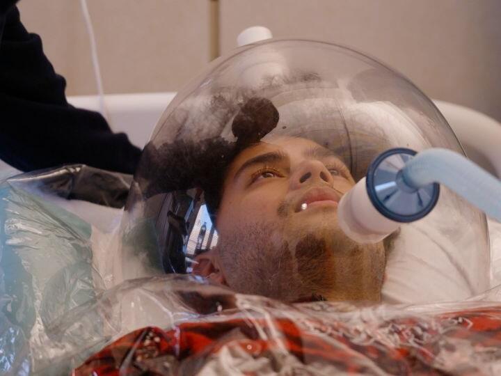
Researchers have developed a device that can simultaneously measure six markers of brain health. The sensor, which is inserted through the skull into the brain, can pull off this feat thanks to an artificial intelligence (AI) system that pieces apart the six signals in real time.
Being able to continuously monitor biomarkers in patients with traumatic brain injury could improve outcomes by catching swelling or bleeding early enough for doctors to intervene. But most existing devices measure just one marker at a time. They also tend to be made with metal, so they can’t easily be used in combination with magnetic resonance imaging.
Simultaneous access to measurements could improve outcomes for brain injuries.

Researchers have developed a new, fast, and rewritable method for DNA computing that promises smaller, more powerful computers.
This method mimics the sequential and simultaneous gene expression in living organisms and incorporates programmable DNA circuits with logic gates. The improved process places DNA on a solid glass surface, enhancing efficiency and reducing the need for manual transfers, culminating in a 90-minute reaction time in a single tube.
Advancements in DNA-Based Computation.
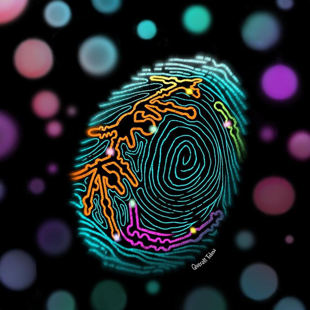
Different types of cancer have unique molecular “fingerprints” which are detectable in early stages of the disease and can be picked up with near-perfect accuracy by small, portable scanners in just a few hours, according to a study published today in the journal Molecular Cell.
The discovery by researchers at the Centre for Genomic Regulation (CRG) in Barcelona sets the foundation for creating new, non-invasive diagnostic tests that detect different types of cancer faster and earlier than currently possible.
The study centers around the ribosome, the protein factories of a cell. For decades, ribosomes were thought to have the same blueprint across the human body. However, researchers discovered a hidden layer of complexity—tiny chemical modifications which vary between different tissues, developmental stages, and disease.