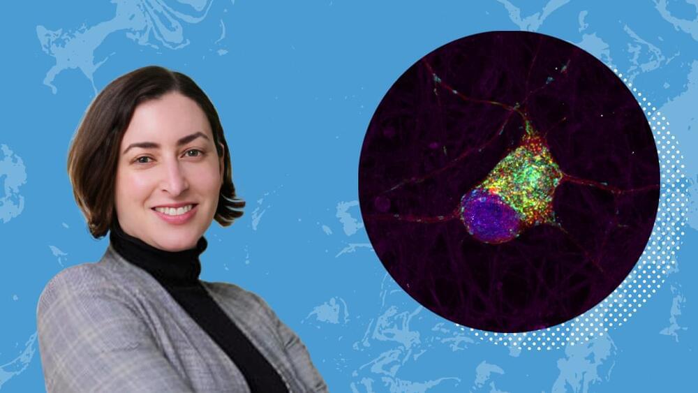
Sarah Cohen and two others will pioneer a method to view these molecular structures and their interactions in live stem cells.

Researchers at the University of Oklahoma have developed a breakthrough method of adding a single nitrogen atom to molecules, unlocking new possibilities in drug research and development. Now published in the journal Science, this research is already gaining international attention from drug manufacturers.
Nitrogen atoms and nitrogen-containing chemical structures, called heterocycles, play a pivotal role in medicinal chemistry and drug development. A team led by OU associate professor Indrajeet Sharma has demonstrated that by using a short-lived chemical called sulfenylnitrene, researchers can insert one nitrogen atom into bioactive molecules and transform them into new pharmacophores that are useful for making drugs.
This process is called skeletal editing and takes inspiration from Sir Derek Barton, the recipient of the 1969 Nobel Prize in Chemistry.

Geophysicists at ETH Zurich are using models of the lower mantle to identify areas where earthquake waves behave differently than previously assumed. This indicates the presence of zones of rocks that are colder, or have a different composition, than the surrounding rocks. This finding challenges our current understanding of the Earth’s plate tectonics—and presents the researchers with a major mystery.
No one can see inside the Earth. Nor can anyone drill deep enough to take rock samples from the mantle, the layer between the Earth’s core and outermost, rigid layer, the lithosphere, or measure temperature and pressure there. That’s why geophysicists use indirect methods to see what’s going on deep beneath our feet.
For example, they use seismograms, or earthquake recordings, to determine the speed at which earthquake waves propagate. They then use this information to calculate the internal structure of the Earth. This is very similar to how doctors use ultrasound to image organs, muscles or veins inside the body without opening them up.
A recent study from the McGovern Institute for Brain Research shows how interests can modulate language processing in children’s brains and paves the way for personalized brain research.
The paper, which appears in Imaging Neuroscience, was conducted in the lab of MIT professor and McGovern Institute investigator John Gabrieli, and led by senior author Anila D’Mello, a recent McGovern postdoc who is now an assistant professor at the University of Texas Southwestern Medical Center and the University of Texas at Dallas.
“Traditional studies give subjects identical stimuli to avoid confounding the results,” says Gabrieli, who is the Grover Hermann Professor of Health Sciences and Technology and a professor of brain and cognitive sciences at MIT. “However, our research tailored stimuli to each child’s interest, eliciting stronger—and more consistent—activity patterns in the brain’s language regions across individuals.”

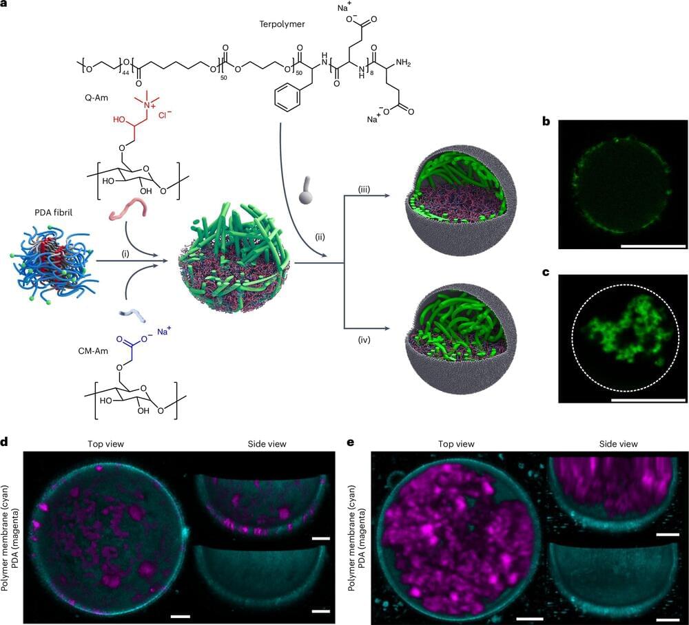
Just like your body has a skeleton, every cell in your body has a skeleton—a cytoskeleton to be precise. This provides cells with mechanical resilience, as well as assisting with cell division. To understand how real cells work, e.g. for drug and disease research, researchers create artificial cells in the laboratory.
However, many artificial cells to date cannot be used to study how cells respond to forces as they don’t have a cytoskeleton. TU/e researchers have designed a polymer-based network for artificial cells that mimics a real cytoskeleton, thus making it possible to study with greater accuracy in artificial cells how cells respond to forces.
The research is published in the journal Nature Chemistry.

Summary: Researchers have developed a Genetic Progression Score (GPS) using artificial intelligence to predict the progression of autoimmune diseases from preclinical symptoms to full disease. The GPS model integrates genetic data and electronic health records to provide personalized risk scores, improving prediction accuracy by 25% to 1,000% over existing models.
This method identifies individuals at higher risk earlier, enabling timely interventions and better disease management. The framework could also be adapted to study other underrepresented diseases, offering a breakthrough in personalized medicine and health equity.

A study from the University of Minnesota Medical School links social stress to accelerated aging, finding that stress damages DNA
DNA, or deoxyribonucleic acid, is a molecule composed of two long strands of nucleotides that coil around each other to form a double helix. It is the hereditary material in humans and almost all other organisms that carries genetic instructions for development, functioning, growth, and reproduction. Nearly every cell in a person’s body has the same DNA. Most DNA is located in the cell nucleus (where it is called nuclear DNA), but a small amount of DNA can also be found in the mitochondria (where it is called mitochondrial DNA or mtDNA).

Scientists have identified a key nucleolar complex that could be instrumental in combating neurodegenerative diseases. This complex plays a critical role in maintaining cellular health by regulating protein homeostasis (proteostasis)—the process by which cells ensure proper protein balance and function.
Research reveals that suppressing this nucleolar complex significantly reduces the toxic effects of proteins associated with Alzheimer’s.
Alzheimer’s disease is a progressive neurological disorder that primarily affects older adults, leading to memory loss, cognitive decline, and behavioral changes. It is the most common cause of dementia. The disease is characterized by the buildup of amyloid plaques and tau tangles in the brain, which disrupt cell function and communication. There is currently no cure, and treatments focus on managing symptoms and improving quality of life.